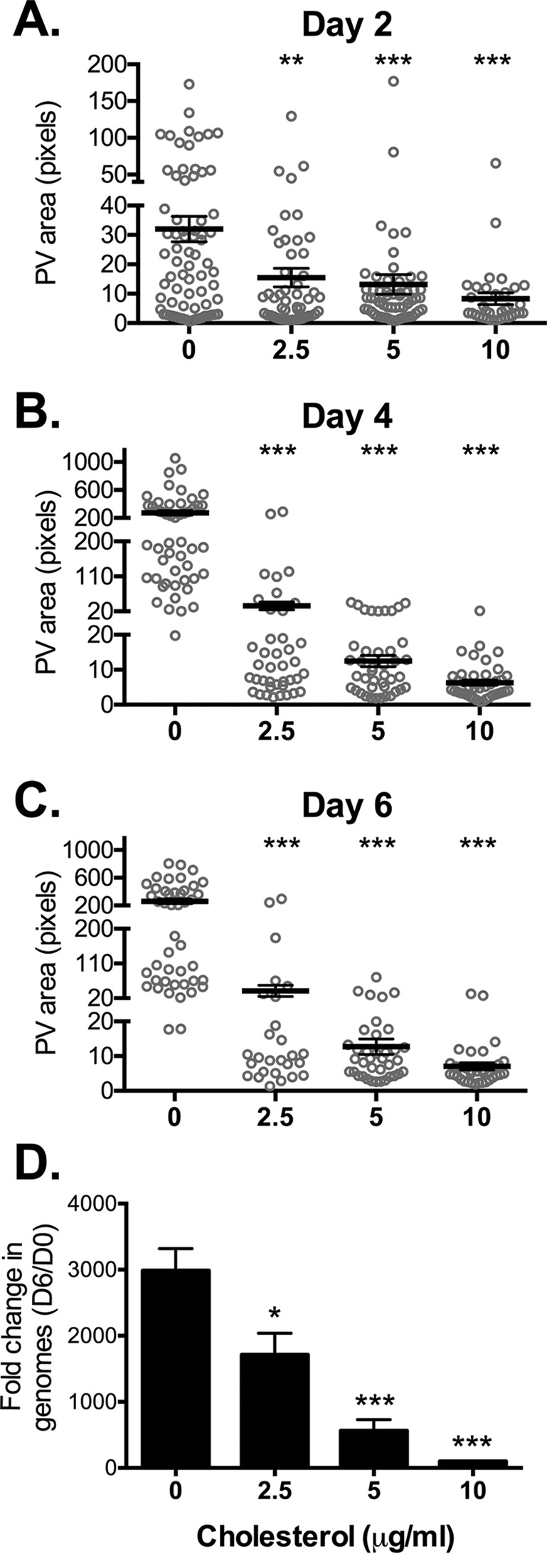
FIG 1 .

PV size and C. burnetii growth are sensitive to cholesterol. (A to C) Measurement of PV sizes reveals that PVs are significantly smaller in MEFs with cholesterol compared to cholesterol-free MEFs. C. burnetii-infected MEFs were incubated with the different cholesterol concentrations (0, 2.5, 5, or 10 µg/ml) and stained by immunofluorescence for C. burnetii and the PV marker LAMP-1 at the indicated times (2, 4, and 6 days). PVs were measured using ImageJ. Each circle represents the value for 1 PV, with at least 15 PVs per condition measured in each of three separate experiments. The means (black horizontal bars) were compared by one-way ANOVA with Tukey’s posthoc test. Error bars represent the standard errors of the means (SEM). The values that were significantly different from the control values (no cholesterol) are indicated by asterisks as follows: **, P < 0.01; ***, P < 0.001. (D) The fold change in bacterial growth under different cholesterol conditions was determined by quantitative PCR for bacterial genomes. The means plus standard deviations (SD) from three separate experiments done in duplicate are shown. Values that are significantly different from the control value (no cholesterol) determined by one-way ANOVA with Dunnett’s posthoc test are indicated by asterisks as follows: *, P < 0.05, ***, P < 0.001. D6, day 6; D0, day 0.
