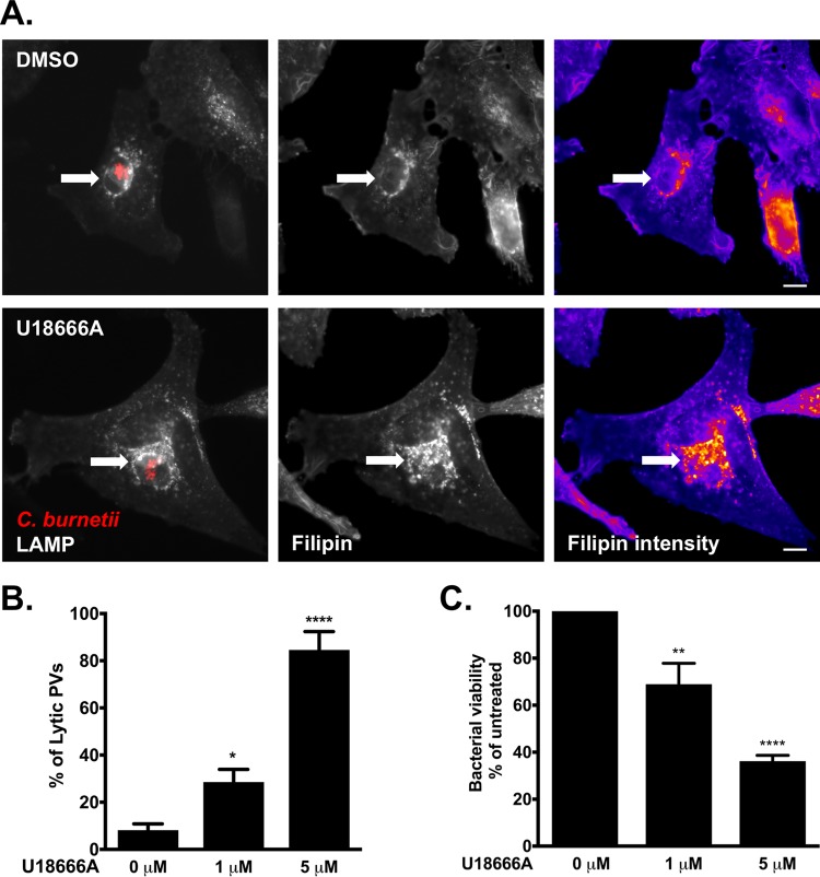
FIG 3 .
Altered cellular cholesterol homeostasis is bactericidal. (A) Microscopy images showing that U18666A treatment traps cholesterol in the C. burnetii PV in HeLa cells. mCherry-C. burnetii-infected HeLa cells were treated with 5 µM U18666A for 6 h, fixed, and stained for sterols (filipin) and PV (LAMP-1). Compared to mock-treated cells, there is an increase in filipin labeling in and around the PV following treatment with U18666A. The white arrows point to the PVs. Filipin intensity is shown as a heat map, with yellow showing the highest filipin intensity and blue showing the lowest filipin intensity. Bars = 5 µm. (B) Quantitation of lytic PVs in U18666A-treated cells after treatment of mCherry-C. burnetii-infected HeLa cells with 1 or 5 µM U18666A. PVs were scored for the presence (lytic) or absence (nonlytic) of free mCherry in the PV lumen, resulting from the lysis of mCherry-expressing bacteria. The means plus SEM (error bars) from three individual experiments are shown. The means were compared to the value with no cholesterol by one-way ANOVA with Dunnett’s posthoc test, and statistically different values are indicated by asterisks as follows: *, P < 0.05; ****, P < 0.0001. (C) C. burnetii viability decreases after 6 h treatment with U18666A. The number of viable bacteria was determined by FFU assay and normalized to the values for the vehicle control (0 µM). The means plus SEM (error bars) from three individual experiments are shown. Statistical significance was determined by comparing values to the value with no cholesterol by one-way ANOVA with Dunnett’s posthoc test and indicated as follows: **, P < 0.01; ****, P < 0.0001. The average values for three independent experiments done in duplicate are shown.

