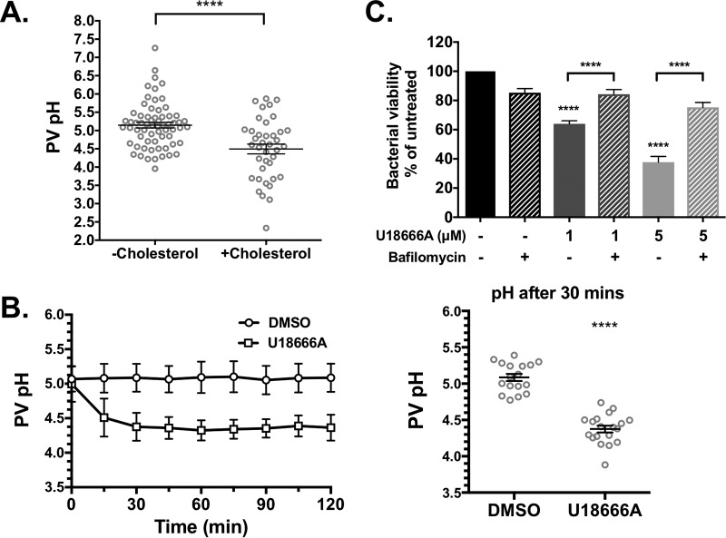
FIG 6 .
Cholesterol accumulation in the PV increases PV acidity. PV pH was determined at 3 days postinfection using a ratiometric fluorescence assay of pH-sensitive Oregon Green dextran and pH-insensitive Alexa 647 dextran. (A) The pH in PVs from MEFs with cholesterol (average pH 4.5) was significantly more acidic than PVs from cholesterol-free MEFs (average pH 5.2). At least 10 PVs were measured in three separate experiments. The means ± SEM (error bars) from three individual experiments are shown. ****, P < 0.0001 as determined by two-tailed unpaired t test. (B) Average PV pH over 2 h in HeLa cells treated with DMSO or 5 µM U18666A. PVs were identified by microscopy and imaged prior to adding drug. While PV pH in DMSO-treated cells remained stable over the time course, PVs in U18666A-treated cells further acidified in the first 30 min. The average pH values at 30 min were 5.1 in DMSO-treated cells and 4.4 in U1666A-treated cells. Approximately 10 PVs were measured in each of two separate experiments, and individual traces are shown in Fig. S4 in the supplemental material. The averages ± SD (error bars) are shown. All time points were significant (P < 0.0001). (C) C. burnetii-infected HeLa cells were treated with U18666A (1 µM or 5 µM) and/or the vATPase inhibitor bafilomycin A1 (100 nM) for 3 h. Bacterial viability, as measured by the FFU assay, is rescued in the presence of bafilomycin. The means plus SEM (error bars) from three individual experiments are shown. ****, P < 0.0001 for the value for U18666A treatment alone compared to the value for no treatment as determined by one-way ANOVA with Tukey’s posthoc test.

