Figure 5.
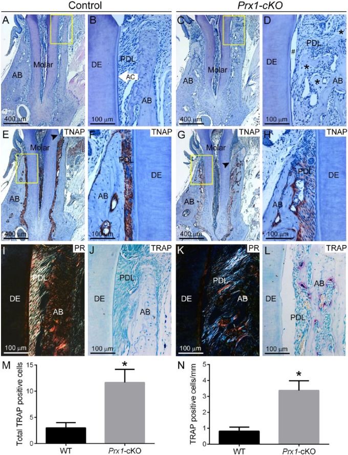
Defective periodontia in Prx1-cKO mice. Dentoalveolar tissues were analyzed by histology in WT control and Prx1-cKO mice (n = 3 per genotype) at 24 wk. By histology, dentoalveolar structures of (A, B) WT molars—including alveolar bone (AB), acellular cementum (AC), periodontal ligament (PDL), and dentin (DE)—are organized and functional. (C, D) Prx1-cKO mice exhibit absence of the acellular cementum layer, PDL detachment (#), osteoid accumulation at the alveolar bone crest (black stars), and invasion of the PDL by alveolar bone. The yellow boxes (A, C) indicate regions of higher magnification (B, D, respectively). Immunohistochemistry for tissue nonspecific alkaline phosphatase (TNAP) in (E, F) WT shows localization in odontoblasts and periodontal cells, whereas (G, H) Prx1-cKO mice exhibit reduced TNAP localized mainly to the bone surface. Black arrowheads (E, G) indicate the location of junctional epithelium, revealing apical migration in Prx1-cKO mice. The yellow boxes (E, G) indicate regions of higher magnification (F, H, respectively). Picrosirius red (PR) staining in (I) WT highlights the highly organized collagen fibers of the PDL, while (K) this selective staining confirms a substantial loss of PDL collagen organization in Prx1-cKO mice. Compared with the few osteoclast-like cells seen in (J) WT alveolar bone, tartrate-resistant acid phosphatase (TRAP) staining in (L) Prx1-cKO mice reveals numerous osteoclast-like cells (large red-purple cells) on alveolar bone surfaces. Quantification and statistical analysis reveals a statistically significant (*P < 0.05) increase in (M) total numbers of TRAP-positive osteoclast-like cells, as well as (N) cells normalized to bone surface, in Prx1-cKO vs. WT mice. cKO, conditional knockout; WT, wild type.

