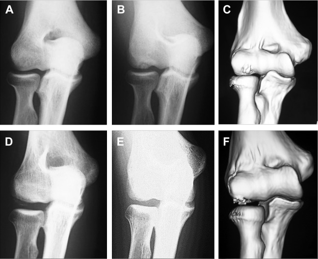
Figure 1.
(A, B, D, and E) Radiographs and (C and F) 3-dimensional computed tomography (3D CT) images of the elbow in (A, B, and C) a 15-year-old baseball player and (D, E, and F) a 16-year-old gymnast with osteochondritis dissecans (OCD) of the humeral capitellum. OCD lesion of the humeral capitellum cannot be detected in the (A) anterior-posterior (AP) view with the elbow in full extension, while the OCD lesion is clearly seen in (B) the AP view with the elbow in 45° of flexion and (C) 3D CT image. On the other hand, OCD lesion is not clearly seen in (E) the AP view with the elbow in 45° of flexion. However, the OCD lesion is clearly seen in (D) the AP image with the elbow in a full extension and (F) 3D CT image.

