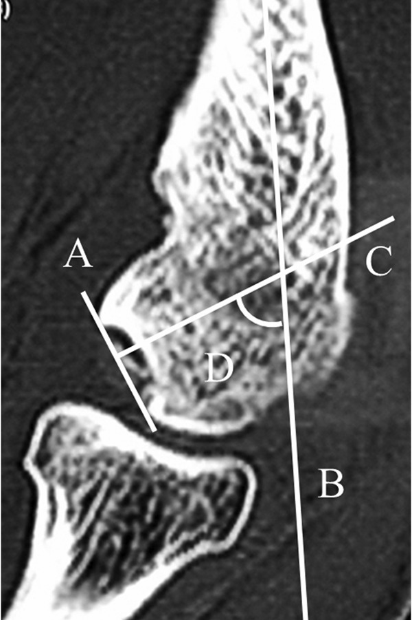
Figure 2.

The inclination angle was measured using a slice that clearly demonstrates the osteochondritis dissecans (OCD) lesion on the sagittal computed tomography image of the elbow. (A) Line connecting the anterior and posterior margin of the OCD lesion; (B) line parallel to the axis of the humerus; (C) line perpendicular to line A; (D) inclination angle, measured as an angle between lines B and C.
