Abstract
Background:
While recent studies have addressed the biomechanical function of the ligamentum teres and provided descriptions of ligamentum teres reconstruction techniques, its detailed quantitative anatomy remains relatively undocumented. Moreover, there is a lack of consensus in the literature regarding the number and morphology of the acetabular attachments of the ligamentum teres.
Purpose:
To provide a clinically relevant quantitative anatomic description of the native human ligamentum teres.
Study Design:
Descriptive laboratory study.
Methods:
Ten human cadaveric hemipelvises, complete with femurs (mean age, 59.6 years; range, 47-65 years), were dissected free of all extra-articular soft tissues to isolate the ligamentum teres and its attachments. A coordinate measuring device was used to quantify the attachment areas and their relationships to pertinent open and arthroscopic landmarks on both the acetabulum and the femur. The clock face reference system was utilized to describe acetabular anatomy, and all anatomic relationships were described using the mean and 95% confidence intervals.
Results:
There were 6 distinct attachments to the acetabulum and 1 to the femur. The areas of the acetabular and femoral attachment footprints of the ligamentum teres were 434 mm2 (95% CI, 320-549 mm2) and 84 mm2 (95% CI, 65-104 mm2), respectively. The 6 acetabular clock face locations were as follows: anterior attachment, 4:53 o’clock (95% CI, 4:45-5:02); posterior attachment, 6:33 o’clock (95% CI, 6:23-6:43); ischial attachment, 8:07 o’clock (95% CI, 7:47-8:26); iliac attachment, 1:49 o’clock (95% CI, 1:04-2:34); and a smaller pubic attachment that was located at 3:50 o’clock (95% CI, 3:41-4:00). The ischial attachment possessed the largest cross-sectional attachment area (127.3 mm2; 95% CI, 103.0-151.7 mm2) of all the acetabular attachments of the ligamentum teres.
Conclusion:
The most important finding of this study was that the human ligamentum teres had 6 distinct points of attachment on the acetabulum (transverse, anterior, and posterior margins of the acetabular notch and cotyloid fossa attachments: ilium, ischium, and pubis) and 1 on the femur. On the acetabulum, the anterior attachment was substantially larger than the posterior attachment and was located at a mean clock face position of 4:53 o’clock.
Clinical Relevance:
These quantitative descriptions of the ligamentum teres can be used by clinicians to arthroscopically identify the attachments of the ligamentum teres, guiding arthroscopic surgical interventions designed to address ligamentum teres pathology.
Keywords: hip, instability, arthroscopy, anatomy
Prior to the advent of hip arthroscopy, surgical diagnosis and treatment of the ligamentum teres were rare because of the necessity to transect the ligamentum teres to gain access to the hip joint.2 However, with the introduction and widespread adoption of arthroscopy as a means for addressing intra-articular hip pathology, renewed attention has been ascribed to potential pathology of the ligamentum teres. Although the ligamentum teres has been described as a vestigial structure due to its slack state during distraction7 and some studies reporting no differences after sectioning the ligamentum teres for surgical dislocation,6 recent studies have reported improved functional and clinical outcomes after arthroscopic debridement or reconstruction of the ligamentum teres.8,11,13,14
Previous studies have analyzed the ligamentum teres to determine its anatomy and biomechanical function. Qualitatively, the ligamentum teres has been reported to have a broad acetabular attachment, with 2 bands attached to the ischial and pubic sides of the acetabular notch that blend with the transverse acetabular ligament.3,9,17 Rao et al14 described the band on the ischial side as stronger and marginally broader, but this observation has not been biomechanically tested or anatomically validated. On its course to the femoral head, the acetabular attachments of the ligamentum teres converge, assuming a ribbon-like shape before attaching into the fovea capitis posteroinferiorly in relation to the center of the femoral head.1
The biomechanical properties of the ligamentum teres were described in a recent study by Martin et al,10 which reported that the ligamentum teres provided restraint to internal and external rotation of the hip in deep hip flexion (>90°). In addition, the ligamentum teres has been described as a secondary hip stabilizer by restricting subluxation in deep flexion, adduction, and external rotation.16 Collectively, the data presented in these studies appear to favor preservation of the ligamentum teres during surgical intervention.
Currently, the literature lacks quantitative anatomic data regarding the attachments of the ligamentum teres on the acetabulum and fovea capitis. Furthermore, there is a lack of consensus in the literature regarding the number of acetabular attachments and their morphology. Therefore, the purpose of this study was to provide a thorough quantitative description of the native ligamentum teres in human cadaveric specimens. Specifically, this study aimed to describe the areas of the attachments of the ligamentum teres and their relation to pertinent open and arthroscopic bony landmarks on the acetabulum and femur.
Methods
Specimen Preparation
Ten fresh-frozen, full human cadaveric pelvises, complete with intact femurs (mean age, 59.6 years; range, 47-65 years; 8 male, 2 female), without osteoarthritis, gross hip morphology abnormality, or surgical history were used in this study. Specimens were stored at −20°C and thawed for 24 hours prior to testing. Specimens were dissected of all extra-articular soft tissues. The cadaveric specimens utilized in this study were donated to a tissue bank for the purpose of medical research and then purchased by our institution.
The femoral head was removed from the acetabulum to gain full exposure of all landmarks. After disarticulation, the femur and pelvis were rigidly clamped in custom fixtures, and a portable coordinate measuring device (7315 Romer Absolute Arm; Hexagon Metrology) was used to quantify the location of pertinent bony landmarks for establishing the femur’s anatomic axes. To identify the location of the center of the femoral head, points encompassing the surface of the femoral head were selected and subjected to a spherical best-fit algorithm.
Quantitative Measurements
With the femur rigidly fixed, pertinent bony and soft tissue landmarks were identified and measured using the coordinate measuring device. The femoral bony and soft tissue landmarks included the femoral attachment of the ligamentum teres, the fovea capitis, and the most superior aspect of the greater trochanter. To establish the axis of the femoral neck, 3 rings of points around the neck were collected (medial, central, and lateral neck rings). Utilizing the same methods, 3 rings of points were collected around the shaft of the femur (proximal, middle, and distal rings).
Next, the bony and soft tissue reference landmarks of interest were identified and collected on each hemipelvis. The bony and soft tissue landmarks on the acetabulum included all acetabular attachments of the ligamentum teres, acetabular rim, stellate crease, psoas-u, transverse acetabular ligament, cotyloid fossa, and the medial cortex.12
Data Analysis
Data were analyzed using custom software (MATLAB 2008b; The MathWorks Inc). Distance measurements were collected as the 3-dimensional linear distance between structures and are referred as direct distances. Unless otherwise noted, all anatomic distance measurements were measured between the centers of the 2 structures. Cross-sectional areas were computed by projecting points taken along the circumference of the attachment onto an interpolated plane and calculating the area of the resulting 2-dimensional polyhedron. Clock face measurements were defined by interpolating a circle from 60 points taken along the acetabular rim. The orientation of the clock face was defined by setting the superior margin of the anterior labral sulcus (psoas-u) at 3 o’clock.12 Left hips were reverted so that they could be considered to have the psoas-u at 3 o’clock to match the right hip measurements. Average measurements across the 10 specimens were reported with 95% confidence intervals (lower bound, upper bound).
Results
Acetabular Attachments
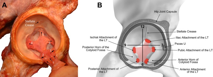
The ligamentum teres had a broad acetabular attachment, with 6 distinct attachment locations (Figure 1). Superficially, the ligamentum teres blended with the transverse acetabular ligament along its entire length (25.0 mm; 95% CI, 22.9-27.0 mm). Beneath the transverse attachment, 2 substantial bands acted to anchor the ligamentum teres to the ischial (posterior) and pubic (anterior) sides of the acetabular notch. The anterior attachment was substantially larger, had a mean cross-sectional attachment area of 79.4 mm2 (95% CI, 53.8-105.0 mm2), and was located at a mean clock face position of 4:53 o’clock (95% CI, 4:45-5:02). The smaller posterior attachment had a mean cross-sectional attachment area of 54.5 mm2 (95% CI, 53.8-105.0 mm2) and was located at a mean clock face position of 6:33 o’clock (95% CI, 6:23-6:43). Superior to these landmarks, there were 3 attachments to each of the 3 bones that converge within the cotyloid fossa (ilium, ischium, and pubis). The ischial attachment possessed the largest cross-sectional attachment area (127.3 mm2; 95% CI, 103.0-151.7 mm2) of all the acetabular attachments of the ligamentum teres and was located at 8:07 o’clock (95% CI, 7:47-8:26), followed closely by the iliac attachment (124.2 mm2; 95% CI, 93.31-155.15), located at a mean location of 1:49 o’clock (95% CI, 1:04-2:34). Finally, a smaller pubic attachment (46.08 mm2; 95% CI, 36.66-55.50 mm2) was located at 3:50 o’clock (95% CI, 3:41-4:00) (Table 1). The areas, clock face positions, and relative distances of these attachments are presented in Tables 1 through 3. The acetabular cortex thickness was also measured at each attachment site (Table 4). The distance to the medial cortex was greatest at the anterior attachment (11.28 mm; 95% CI, 9.09-13.46 mm).
Figure 1.
(A) Photograph and (B) illustration of the ligamentum teres (LT) attachment locations on the acetabulum. 1 = transverse attachment (to the transverse acetabular ligament, on the reverse side of the ligamentum teres from what is shown); 2 = posterior attachment; 3 = anterior attachment; 4 = ischial attachment; 5 = iliac attachment; 6 = pubic attachment.
TABLE 1.
Area Measurements of the Acetabular Attachments of the Ligamentum Teresa
| Attachment | Mean Area | 95% CI, Low | 95% CI, High |
|---|---|---|---|
| Superficial attachment (transverse acetabular ligament length), mm | 24.95 | 22.88 | 27.02 |
| Anterior attachment | 79.36 | 53.76 | 104.95 |
| Posterior attachment | 54.49 | 36.10 | 72.88 |
| Ischial attachment | 127.34 | 102.99 | 151.69 |
| Iliac attachment | 124.23 | 93.31 | 155.15 |
| Pubic attachment | 46.08 | 36.66 | 55.50 |
| Total acetabular attachment | 431.49 | 361.77 | 501.22 |
aValues are presented as mm2 unless otherwise noted.
TABLE 2.
Clock Face Positions of the Acetabular Attachments of the Ligamentum Teres
| Attachment | Clock Face Position | 95% CI, Low | 95% CI, High |
|---|---|---|---|
| Transverse acetabular ligament pubic attachment | 4:39 | 4:32 | 4:46 |
| Transverse acetabular ligament ischial attachment | 6:30 | 6:22 | 6:39 |
| Anterior attachment | 4:45 | 4:42 | 4:49 |
| Posterior attachment | 6:25 | 6:15 | 6:34 |
| Ischial attachment | 7:58 | 7:39 | 8:17 |
| Iliac attachment | 1:46 | 1:01 | 2:30 |
| Pubic attachment | 3:42 | 3:36 | 3:49 |
TABLE 3.
Direct Distances From the Acetabular Attachments of the Ligamentum Teres to the Center of the Clock Face
| Attachment | Mean Distance, mm | 95% CI, Low | 95% CI, High |
|---|---|---|---|
| Anterior attachment | 25.05 | 23.89 | 26.21 |
| Posterior attachment | 24.83 | 22.88 | 26.79 |
| Ischial attachment | 11.83 | 10.11 | 13.54 |
| Iliac attachment | 6.79 | 5.11 | 8.47 |
| Pubic attachment | 16.94 | 14.96 | 18.92 |
TABLE 4.
Thickness of Acetabular Bone at Each Attachment Site of the Ligamentum Teres
| Attachment | Cortex Thickness, mm | 95% CI, Low | 95% CI, High |
|---|---|---|---|
| Anterior attachment | 11.28 | 9.09 | 13.46 |
| Posterior attachment | 6.79 | 5.01 | 8.57 |
| Ischial attachment | 6.00 | 4.66 | 7.34 |
| Iliac attachment | 7.76 | 6.24 | 9.27 |
| Pubic attachment | 6.62 | 4.32 | 8.92 |
Femoral Attachments
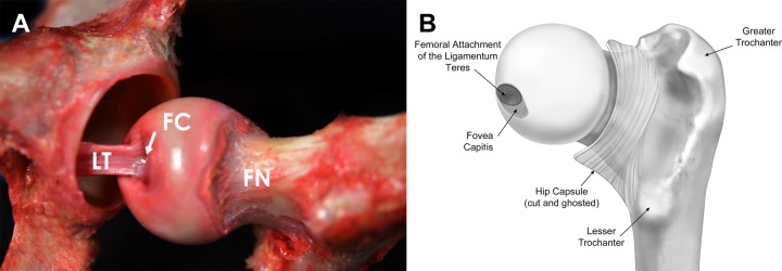
As the ligamentum teres coursed distally toward the femoral head, there was a confluence of its fibers to a single location within the anterosuperior region of the fovea capitis (Figure 2). The femoral attachment of the ligamentum teres had an oval shape with a mean area of 84.40 mm2 (95% CI, 64.90-103.90 mm2), which comprised approximately 43.38% (95% CI, 37.76%-48.99%) of the area of the fovea capitis.
Figure 2.
(A) Photograph and (B) illustration of the attachment location of the ligamentum teres (LT) on the femur. FC, fovea capitis; FN, femoral neck.
The distance from the superior-most tip of the greater trochanter to the femoral attachment of the ligamentum teres was 12 mm (95% CI, 5.83-20.10 mm) anterior, 1.51 mm (95% CI, −1.07 to 4.09 mm) superior, and 66.78 mm (95% CI, 62.43-71.14 mm) medial. The direct distance from the superior tip of the greater trochanter to the femoral attachment of the ligamentum teres was 69.16 mm (95% CI, 65.80-72.52 mm).
Discussion
The most important finding of this study was that the ligamentum teres consistently attached at 6 acetabular locations and 1 femoral location in all human cadaveric specimens analyzed. On the acetabular side, the ligamentum teres had a pyramidal shape1,3 with unique bands anchoring it to 6 distinct locations. The first band, previously referred to as the medial bundle,5 attached along the entire length of the transverse acetabular ligament. In addition, the ligamentum teres had 2 bands with bony attachments to the ischial and pubic margins of the acetabular notch. Finally, there were 3 attachments to each of the 3 bones that converge within the cotyloid fossa (ilium, ischium, and pubis), located at the margin of the cotyloid fossa and articular cartilage. As the ligamentum teres coursed toward its femoral attachment at the fovea capitis, there was a confluence of its ribbon-like fibers into a compact, tubular ligamentous structure. The convergence of 6 attachments into 1 femoral attachment presents increased complexity for reconstruction procedures that attempt to re-create the native anatomy by selecting an appropriately sized graft and fixing it in anatomic locations. Current arthroscopic ligamentum teres reconstructions focus on reproducing only 1 attachment location for both the acetabulum and femur, as it is less technically demanding than reconstructing each bundle.
The current study both confirms and further expands on the reported qualitative attachment locations of the ligamentum teres in previous studies. Generally, the ligamentum teres has been described as attaching broadly to the cotyloid fossa,1 with 2 distinct bands originating from the ischial and pubic regions of the acetabular notch. These fibrous bands blend anteroinferiorly with the transverse ligament.3,9,17 As the ligamentum teres courses to its femoral head attachment, the ligament assumes an oval shape and inserts at the fovea capitis, which typically lies posterior and inferior to the femoral head center.1 Demange et al5 described 3 constituent bands of the ligamentum teres: posterior, anterior, and medial; however, these descriptions were not verified by the literature. The ligamentum teres is reported to be variable in length and absent in 10% of individuals.15 The ligamentum teres has a mean cross-sectional length between 30.6 and 59 mm2.12
The ligamentum teres was previously considered a vestigial structure, lacking a pivotal role in the function of the hip.7 However, recent studies propose that the ligamentum teres has a more significant role than previously recognized.11,13,14 Rao et al14 described the morphology and structural properties of the attachments of the ligamentum teres, concluding that the posterior attachments were stronger and marginally broader. In an assessment of the role of the ligamentum teres in providing range of motion stability, Martin et al10 reported significant increases in internal rotation and external rotation when the hip was in 90° or 120° of flexion after ligamentum teres sectioning. A similar finding was reported by van Arkel et al,16 confirming that the ligamentum teres was a secondary restraint in high flexion, adduction, and external rotation. The ligamentum teres has a small role in limiting external rotation but its contribution is considerably less than that of the lateral iliofemoral ligament, indicating that repair of the ligamentum teres to restore native restraint should only be considered secondary to repair of any deficiencies in the anterior joint capsule. During anatomic dissection in the current study, the posterior and anterior bands tightened during internal and external rotation, respectively. This structural design, with 2 anchoring points at 5:00 and 6:30, may afford increased ability for the ligamentum teres to restrain end-range rotational movements compared with a single attachment location. Therefore, a reconstruction of the ligamentum teres may need to replicate these 2 attachment locations.
A recent study reported that among 2213 patients with femoroacetabular impingement and labral pathology, complete tears of the ligamentum teres were found in 1.5% of patients and were more likely to be seen in women, patients with a lower body mass index, and patients with low center-edge angles.4 Ligamentum teres tears were associated with increased hip laxity and chondral defects of the femoral head.4 The role of the ligamentum teres may be especially important in patients with dysplasia of the hip or joint hypermobility because deficiency of the normal bony architecture and/or soft tissue restraints about the hip can lead to increased reliance on the ligamentum teres as a secondary stabilizer.10 Therefore, these abnormalities in a patient with a ligamentum teres tear more commonly were indicative of a need for a ligamentum teres reconstruction in addition to treatment of the underlying pathology.10,13
We recognize that the present study has some limitations inherent to a cadaveric study design. The analysis was performed through an open approach, which did not have the distraction or pressure distension present during arthroscopy, which may have altered the appearance of anatomic structures. Although a meticulous dissection was performed to clearly visualize the ligaments, fiber direction, and attachment sites, distances were calculated as absolute 2-dimensional distances, which may have resulted in underestimation of structure lengths due to out-of-plane length contributions along the course of the ligament measured. Also, due to the nature of this study, we did not measure femoral torsion. This could possibly influence the distance of the femoral attachment of the ligamentum teres to the other landmarks on the femur. Finally, this study was performed on 10 cadaveric hemipelvises and may have not included some of the anatomic variability seen in the greater population.
Conclusion
The most important finding of this study was that the human ligamentum teres had 6 distinct points of attachment on the acetabulum (transverse, ischial, and pubic margins of the acetabular notch and cotyloid fossa attachments: ilium, ischium, and pubis) and 1 on the femur. On the acetabulum, the anterior attachment was substantially larger than the posterior attachment and was located at a mean clock face position of 4:53 o’clock.
Acknowledgment
The authors would like to thank David M. Civitarese, BA, for his assistance with specimen and supply acquisition and organization, Bradley M. Kruckeberg, BA, for his assistance with data collection, and Andy Evansen for his expertise in creatively developing medical illustrations.
Footnotes
One or more of the authors has declared the following potential conflict of interest or source of funding: R.F.L. receives royalties from Arthrex and Smith & Nephew; is a paid consultant for Arthrex, Ossur, and Smith & Nephew; and receives research support from Arthrex, Smith & Nephew, Ossur, and Linvatec. M.J.P. receives royalties from Conmed Linvatec, Smith & Nephew, Bledsoe, Donjoy, and Arthrosurface; is a paid consultant for Smith & Nephew; holds patents with Smith & Nephew; is a stockholder in Arthrosurface, MJP Innovations LLC, and MIS; and receives research support from Ossur, Siemens, Smith & Nephew, and Vail Valley Medical Center. This study was internally funded by the Steadman Philippon Research Institute.
Ethical approval was not sought for the present study.
References
- 1. Bardakos NV, Villar RN. The ligamentum teres of the adult hip. J Bone Joint Surg Br. 2009;91:8–15. [DOI] [PubMed] [Google Scholar]
- 2. Byrd JW, Jones KS. Traumatic rupture of the ligamentum teres as a source of hip pain. Arthroscopy. 2004;20:385–391. [DOI] [PubMed] [Google Scholar]
- 3. Cerezal L, Kassarjian A, Canga A, et al. Anatomy, biomechanics, imaging, and management of ligamentum teres injuries. Radiographics. 2010;30:1637–1651. [DOI] [PubMed] [Google Scholar]
- 4. Chahla J, Soares EA, Devitt BM, et al. Ligamentum teres tears and femoroacetabular impingement: prevalence and preoperative findings. Arthroscopy. 2016;32:1293–1297. [DOI] [PubMed] [Google Scholar]
- 5. Demange MK, Kakuda CMS, Pereira CAM, Sakaki MH, Albuquerque RFM. Influence of the femoral head ligament on hip mechanical function. Acta Ortop Bras. 2007;15:187–190. [Google Scholar]
- 6. Ganz R, Gill TJ, Gautier E, Ganz K, Krugel N, Berlemann U. Surgical dislocation of the adult hip a technique with full access to the femoral head and acetabulum without the risk of avascular necrosis. J Bone Joint Surg Br. 2001;83:1119–1124. [DOI] [PubMed] [Google Scholar]
- 7. Gray AJ, Villar RN. The ligamentum teres of the hip: an arthroscopic classification of its pathology. Arthroscopy. 1997;13:575–578. [DOI] [PubMed] [Google Scholar]
- 8. Haviv B, O’Donnell J. Arthroscopic debridement of the isolated ligamentum teres rupture. Knee Surg Sports Traumatol Arthrosc. 2011;19:1510–1513. [DOI] [PubMed] [Google Scholar]
- 9. Hosalkar HS, Varley ES, Glaser D, Farnsworth CL, Bomar JD, Wenger DR. Isocentric reattachment of ligamentum teres: a porcine study. J Pediatr Orthop. 2011;31:847–852. [DOI] [PubMed] [Google Scholar]
- 10. Martin HD, Hatem MA, Kivlan BR, Martin RL. Function of the ligamentum teres in limiting hip rotation: a cadaveric study. Arthroscopy. 2014;30:1085–1091. [DOI] [PubMed] [Google Scholar]
- 11. Philippon M, Schenker M, Briggs K, Kuppersmith D. Femoroacetabular impingement in 45 professional athletes: associated pathologies and return to sport following arthroscopic decompression. Knee Surg Sports Traumatol Arthrosc. 2007;15:908–914. [DOI] [PMC free article] [PubMed] [Google Scholar]
- 12. Philippon MJ, Michalski MP, Campbell KJ, et al. An anatomical study of the acetabulum with clinical applications to hip arthroscopy. J Bone Joint Surg Am. 2014;96:1673–1682. [DOI] [PubMed] [Google Scholar]
- 13. Philippon MJ, Pennock A, Gaskill TR. Arthroscopic reconstruction of the ligamentum teres: technique and early outcomes. J Bone Joint Surg Br. 2012;94:1494–1498. [DOI] [PubMed] [Google Scholar]
- 14. Rao J, Zhou YX, Villar RN. Injury to the ligamentum teres. Mechanism, findings, and results of treatment. Clin Sports Med. 2001;20:791–799. [DOI] [PubMed] [Google Scholar]
- 15. Tan CK, Wong WC. Absence of the ligament of head of femur in the human hip joint. Singapore Med J. 1990;31:360–363. [PubMed] [Google Scholar]
- 16. van Arkel RJ, Amis AA, Cobb JP, Jeffers JR. The capsular ligaments provide more hip rotational restraint than the acetabular labrum and the ligamentum teres: an experimental study. Bone Joint J. 2015;97-B:484–491. [DOI] [PMC free article] [PubMed] [Google Scholar]
- 17. Wenger D, Miyanji F, Mahar A, Oka R. The mechanical properties of the ligamentum teres: a pilot study to assess its potential for improving stability in children’s hip surgery. J Pediatr Orthop. 2007;27:408–410. [DOI] [PubMed] [Google Scholar]


