Fig. 3.
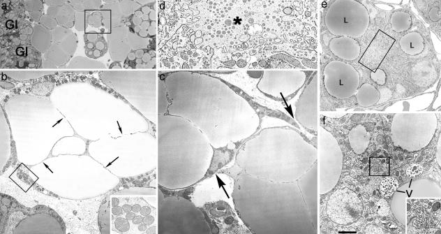
During pregnancy, adipocytes of the mammary gland exhibit epithelial features. Mouse mammary gland on day 18 of pregnancy. (a) Light microscopy. (b–f) Electron microscopy. (a) Adipocytes near the newly formed alveoli (Gl) show lipid droplet compartmentalization. (b) Electron microscopy of the area squared in a. Adipocytes show a thickened peripheral cytoplasm containing unusually large mitochondria (enlarged in Inset) and thin cytoplasmic processes (arrows) that seem to subdivide the lipid droplet. (c) Transdifferentiating adipocytes also exhibit cytoplasmic projections (arrows), suggesting a tendency to become joined and to give rise to early alveolar structures (see d–f). (d) Enlargement of the boxed area in e, showing a small lumen (asterisk) with microvilli. (e) Early alveolar structure containing large lipid droplets (L). (f) Vacuoles (V) containing milk protein secretory granules are present in some early alveolar structures (see the lumen crowded with microvilli in the boxed area, which is enlarged in Inset). [Scale bar: 15 (a), 2.5 (b), 0.5 (b Inset), 2.5 (c), 0.5 (d), 2 (e), 1.2 (f), and 0.4 (f Inset) μm.]

