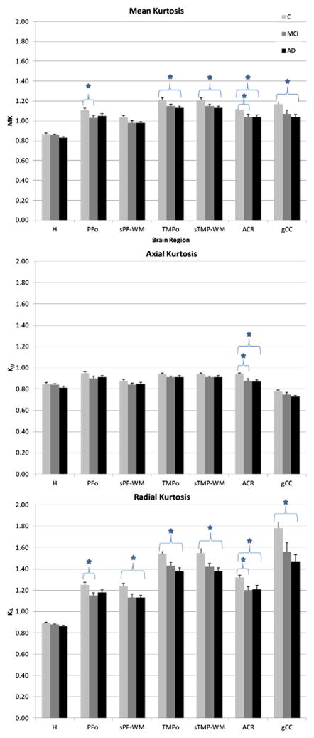
Fig. 3.
Means (+/− s.d.) for kurtosis metrics in each ROI, for each study group. Indices that were found to be statistically significant after Tukey’s multiple comparison correction (significance defined as p < 0.05) are labeled with *. ROIs: segmented prefrontal white matter (sPF-WM); prefrontal oval (PFo); genu of the corpus callosum (gCC); anterior corona radiata (ACR); segmented temporal white matter (sTMP-WM); temporal oval (TMPo); hippocampus (H). C = controls; MCI = mild cognitive impairment; AD = Alzheimer’s disease.

