
Figure 1. Identification of carotid plaque characteristics on multi-contrast MRI.
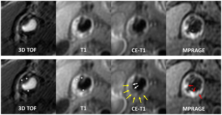
Upper panel: multicontrast images of a representative carotid plaque with all the common plaque characteristics. Lower panel: markers for specific plaque characteristics are added. Areas that are hypointense on all contrast weightings indicate calcification (asterisks); non-calcified areas that have no- or little-enhancement on post-contrast images indicate LRNC (yellow arrows); hyperintense signals on MPRAGE indicate IPH (red arrows). The absence of fibrous cap signals (LRNC is immediately adjacent to lumen) on TOF and CE-T1W indicates the presence of thin/ruptured fibrous cap (white arrows). CE-T1W = contrast enhanced T1-weighted; LRNC = lipid-rich necrotic core; T1W = T1-weighted; T2W = T2-weighted; TOF = time-of-flight.