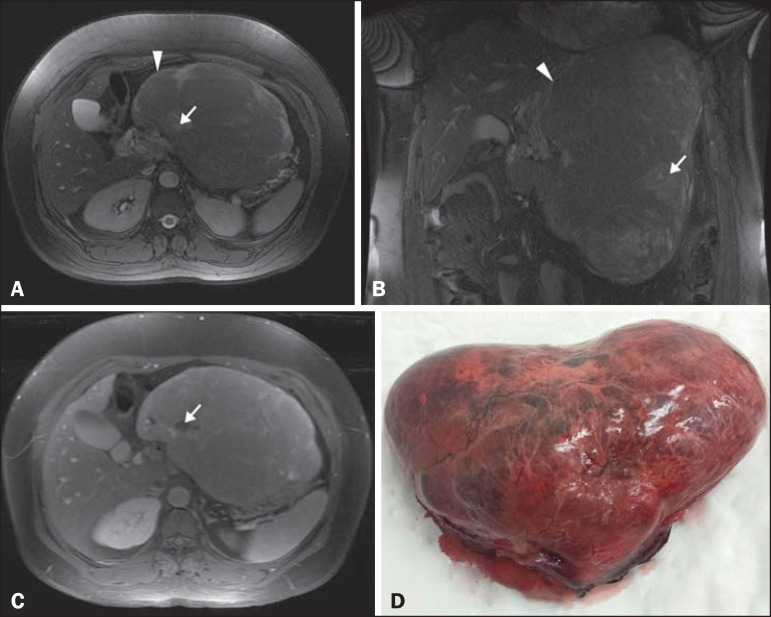
Figure 1.
A,B: Axial and coronal fast imaging employing steady-state acquisition MRI with fat suppression. Solid, encapsulated, heterogeneous expansive mass in the epigastrium (arrowhead), with lobulated contours and areas of hyperintensity (arrow) consistent with necrosis. C: T1-weighted MRI acquisition with fat suppression after intravenous administration of paramagnetic contrast. Diffuse, heterogeneous paramagnetic contrast uptake by the neoplasm. Note the areas without uptake, which is consistent with necrosis (arrow). D: Macroscopic examination. Specimen received in formalin, designated as the product of a left hepatectomy, consisting of a liver fragment weighing 2354 g and measuring 23 × 17 × 11 cm, with an irregular shape, a smooth, brownish external surface, and a bloody area that measured 10 × 6 cm.

