Fig. 4.
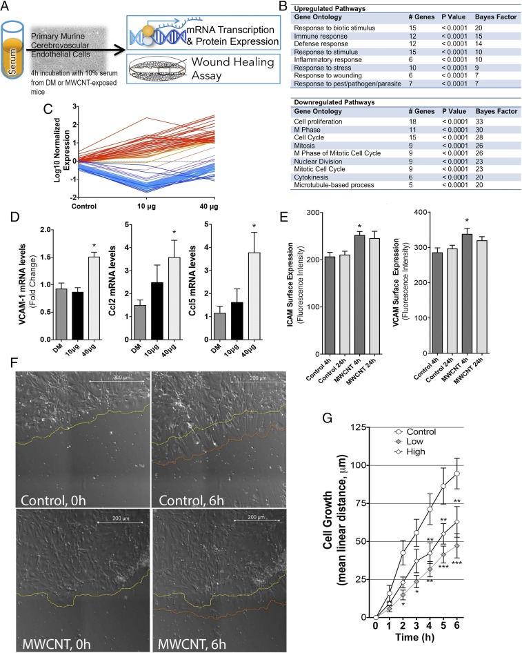
Inflammatory responses of cerebrovascular endothelial cells treated with serum from MWCNT-exposed mice. (A) General depiction of inflammatory potential assay and wound healing protocols, with serum from exposed mice incubated on primary murine cerebrovascular endothelial cells. (B) Microarray results indicating numerous significantly altered transcripts (n = 84) from endothelial cells incubated with serum from MWCNT-exposed mice, compared with cells incubated with serum from control mice (at 4 h after aspiration). (C) Elevated transcripts (in red) were ontologically related to inflammatory and/or cellular defense response, whereas down-regulated transcripts (blue) were related to proliferation and migration pathways, listed in the table. (D) Confirmation of key inflammatory mRNA responses by PCR showing that endothelial Vcam1, Ccl2, and Ccl5 mRNA were significantly up-regulated by serum from MWCNT-treated mice. (E) Endothelial cell surface Icam-1 and Vcam-1 protein expression was elevated by serum from mice exposed to the 40 μg dose of MWCNTs at 4 h compared with controls. *P < 0.05 compared with DM control serum. (F) Live cell images of wound recovery in mCECs treated with serum from control and 40 µg MWCNT-treated mice, showing the initial edge of endothelial cells (yellow dashed line) and the edge after 6 h (orange dashed line). (G) Mean cell regrowth following wounding in primary cerebrovascular endothelial cells incubated with serum from DM- and MWCNT-exposed mice. *P < 0.05; **P < 0.01; ***P < 0.001 compared with DM control serum effects, two-way repeated-measures ANOVA.

