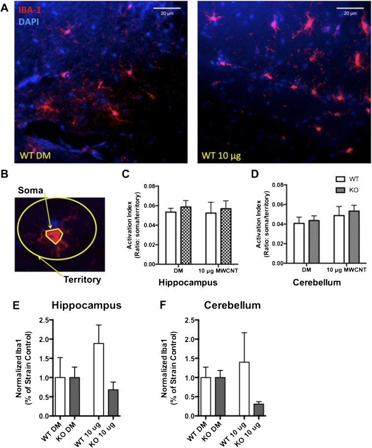
Fig. S2.
Exposure to 10 μg of MWCNTs does not induce microglial activation broadly throughout the brain. Activation of microglia was assessed at 24 h after exposure to 10 μg of MWCNTs in WT and KO animals. (A) Representative images obtained with a 40× objective of the hippocampus for DM WT animals (Left) and WT 10 μg MWCNT-exposed animals (Right). Iba-1 immunohistochemistry (red) indicates microglial cells in this region. (B) Microglial activation was determined by assessing the perimeter of the soma relative to the territory occupied by the ramified processes for each cell and is presented as the ratio of soma to territory. (C and D) Exposure to 10 μg of MWCNTs did not alter the activation index of microglia in the hippocampus (C) or in the cerebellum (D) of animals, as assessed by two-way ANOVA. (E and F) Total Iba-1 immunofluorescence, suggesting that whereas microglia proximal to MWCNT-affected neurovascular units are responding to BBB deficits, the overall population of microglia does not appear to be activated.

