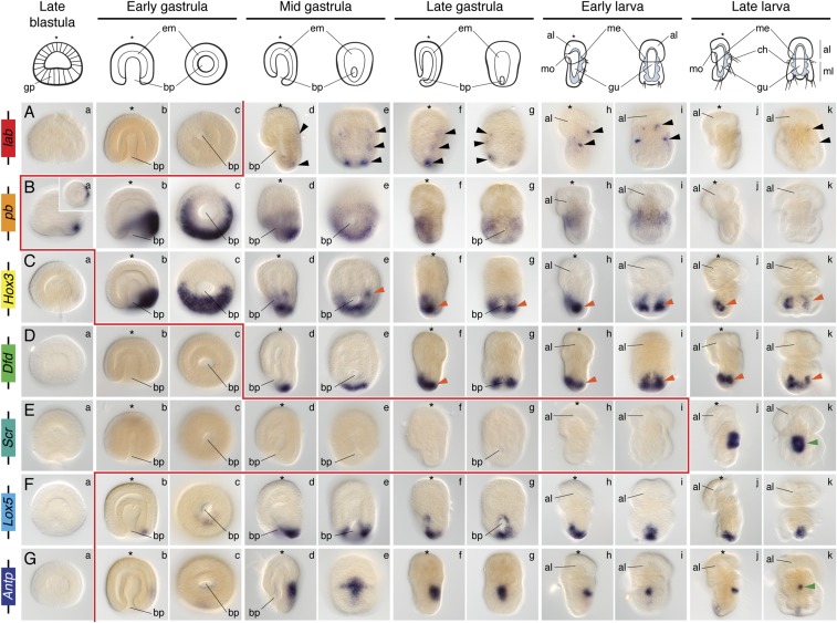
Fig. 4.
Whole-mount in situ hybridization of the Hox genes during embryonic and larval stages in N. anomala. The gene lab is expressed in the chaetae. The Hox genes Hox3 and Dfd are expressed collinearly in the mantle mesoderm. The genes Scr and Antp are expressed in the prospective shell-forming epithelium. The genes pb and Lox5 are detected in the ectoderm of the mantle lobe. The genes Lox4 and Post2 were not detected in transcriptomes and cDNA during embryonic stages. These expression patterns are described in detail in the text. Black arrowheads indicate expression in the chaetae sacs. Orange arrowheads highlight mesodermal expression. Green arrowheads indicate expression in the periostracum. On top are schematic representations of each analyzed developmental stage on its respective perspective. In these schemes, the blue area represents the mesoderm. Drawings are not to scale. The red line indicates the onset of expression of each Hox gene based on in situ hybridization data. The blastula stage is a lateral view (Inset in Ba is a vegetal view). For each other stage, the left column is a lateral view and the right column is a dorsoventral view. The asterisk demarcates the animal/anterior pole. al, apical lobe; bp, blastopore; ch, chaetae; em, endomesoderm; gp, gastral plate; gu, gut; me, mesoderm; ml, mantle lobe; mo, mouth.

