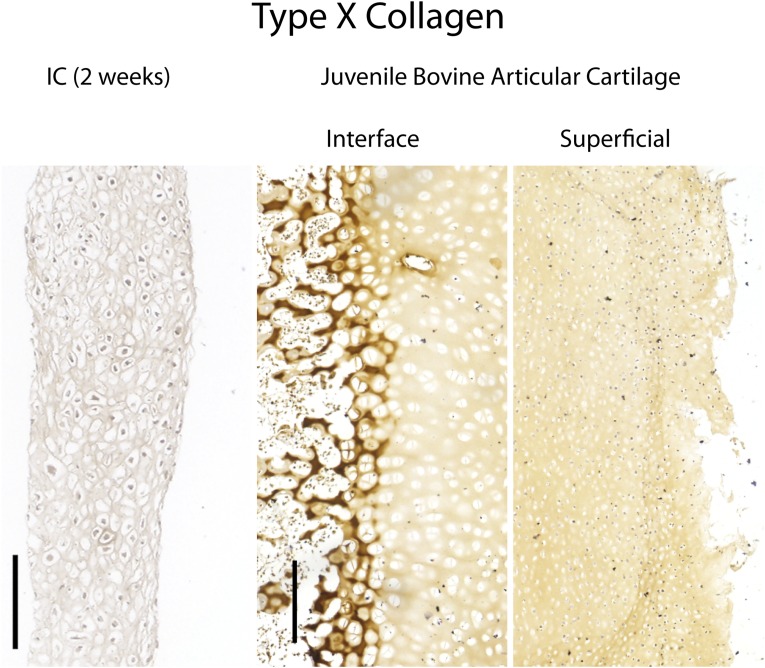
Fig. S7.
Control stains for type X collagen. (Left) Native juvenile (2-mo) osteochondral tissue harvested from a bovine femoral condyle expressed type X collagen at the cartilage–bone interface but not the superficial zone. (Right) Engineered tissues subjected to chondrogenic induction for 2 wk did not express type X collagen. (Scale bars, 200 µm.)

