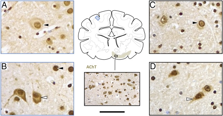
Fig. 4.
Immunohistochemical DIRAS1 expression. Wild-type RRs show predominantly nuclear staining (black arrowhead) as seen in the parietal cortex (A; blue frame) and cholinergic forebrain nuclei (C; black frame). With DIRAS1 mutation (B and D) protein expression is abundant and there is a more diffuse staining of nerve cell perikarya (white arrowhead) in all brain regions, including the brainstem. Figure shows expression in parietal cortex (B; blue frame), and forebrain nuclei (D; black frame). Cholinergic target areas were confirmed by staining for the vesicular acetylcholine transporter (AChT), as demonstrated in the Inset. (Scale bar: A–D, 35 μm; inlet AChT, 150 μm.)

