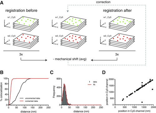
Fig. S1.
Mechanical shift correction for dual-color localization microscopy. (A) Schematic representation of the super registration procedure for dual-color wide-field microscopy used to correct for microscope instability. In addition to the chromatic aberration correction, images were also corrected for mechanical shifts using an average displacement measurement calculated before and after image acquisition. Subdiffraction fluorescent beads were imaged through z stacks in Cy5 (green) and Cy3 (red) channels in between the registration of beads that were imaged in the same wavelengths (before and after registration). Localization of the center of each spectrally separated PSF was determined by a Gaussian fit using FISH_QUANT software (20); all centroids were segregated by pairs, and their distances were measured using MATLAB custom algorithms. (B) Percentage of colocalization between centroids before (black line) and after (red line) correction was applied to the entire FOV. (C) Distribution of observed distances of centroid pairs in two-color images after correction. Data are shown as gray bars, and Gaussian fit is the red line. Mean of distribution = 20.45 ± 0.22 nm. Error, SEM. (D) Scatterplot shows equidistant positions between localized centroids in Cy5 and Cy3 channels.

