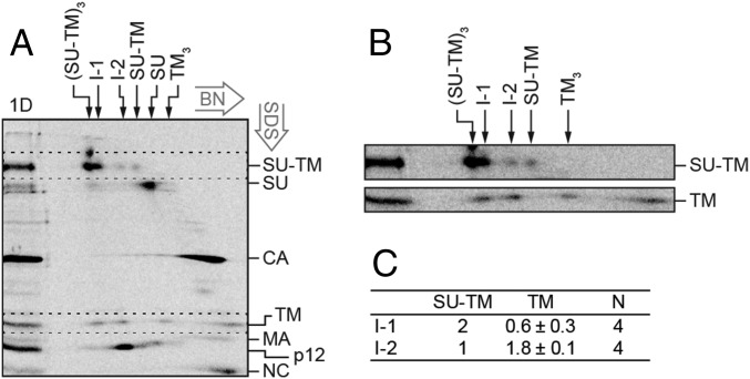
Fig. 3.
2D BN/SDS/PAGE analysis of the compositions of the in vitro activated Env intermediates. [35S] Met/Cys-labeled Mo-MLV was activated as described in Fig. 2 and subjected to BN-PAGE in a first dimension and then to nonreducing SDS/PAGE in a second dimension (A). In the marker lane, to the left, a portion of the sample has been analyzed directly by SDS/PAGE (1D). This provides markers for the migration of the viral proteins in the 2D analysis. The migrations of the viral proteins are indicated to the right, and those of the Env related oligomers in the first dimension BN-PAGE are indicated at the top. The directions of the 2D electrophoreses are indicated. (B) Enlarged cutouts of the SU-TM and TM regions (dotted rectangles) of the 2D gel, at higher contrast. Note the alignment of the two leftmost TM spots with the SU-TM spots of I-1 and I-2. (C) Quantification of the stoichiometric ratio between SU-TM complexes and noncovalently associated TM in I-1 and I-2.

