Fig. 5.
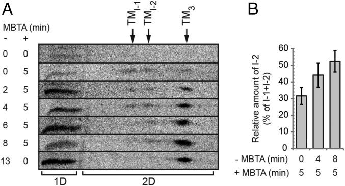
Time-resolved ratio of receptor-activated and MBTA-alkylated Env intermediates. XC cells were spinoculated with [35S] Met/Cys-labeled virus in the cold, shifted to 29 °C, and first incubated for 0–13 min without alkylator (−MBTA) and then in the presence of 0.4 mM MBTA for 0 or 5 min (+MBTA) as indicated, solubilized in the presence of EDTA and NEM, and the virus/cell samples analyzed on 2D BN/SDS/PAGE. (A) Stack of horizontal cutouts from the 2D gels, covering the entire TM region. The positions of I-1, I-2, and TM3, in the first dimension BN-PAGE are indicated at the top. In the cutouts, the noncovalently associated TM of I-1 and I-2 are seen as separate spots to the left of the TM trimer spot. Note the shift in their relative amounts with time of incubation before the alkylation pulse. To the left of the 2D analyses (2D) is TM from samples analyzed by direct SDS/PAGE in the marker lane (1D). (B) Mean relative amount of alkylated I-2, as percentage of the sum of alkylated I-1 and I-2, in samples that have been alkylation pulsed for 5 min at the beginning (after 0 min), in the middle (after 4 min), and at the end (after 8 min) of an activation incubation (± SD; n = 5).

