Fig. S6.
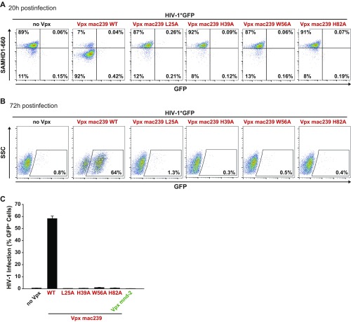
MDMs were infected with VSV-G pseudotyped HIV-1*GFP with incorporated SIVmac239 WT or the depicted mutants using identical TZM-bl infectious units. (A and B) Primary FACS dot plots for SAMHD1 expression and GFP of MDMs from one donor 20 h postinfection (A) and 72 h postinfection (B). (C) Quantification of the percentage of GFP+ cells 72 h postinfection. Depicted is the arithmetic mean + SEM of four donors, each measured in technical duplicates.

