Fig. 1.
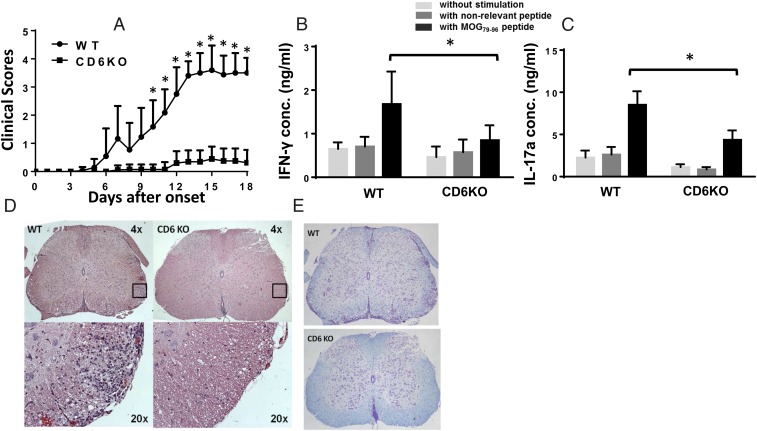
CD6 KO mice are protected from EAE. (A) Clinical scores of the WT and CD6 KO mice. Combined results from four experiments. n = 15 in each group. *P < 0.01. (B) MOG-specific Th1 and Th17 recall assays. Splenocytes from WT and CD6 KO mice 21 d after immunization were incubated without peptide (white bars), with 10 µg/mL of a nonrelevant peptide (IRBP1–20, gray bars) or with 10 µg/mL MOG79–96 peptide (black bars) for 72 h. IFN-γ and IL-17a levels in the culture supernatants were measured by ELISA (n = 14 in each group, *P < 0.05). (C) Spinal cord in CD6 KO mice had markedly reduced leukocyte infiltration as assessed by H&E staining. (D) Representative images of spinal cord sections from the EAE mice after H&E staining, showing that CD6 KO mice have significantly reduced cell infiltration. (Magnification: Upper, 4x; Insets shown at Lower, 20×.) (E) Representative images of spinal cord sections from the EAE mice after Luxol Blue staining, showing that CD6 KO mice had intact myelin sheath (blue staining) compared with severe demyelination in the WT mice. (Magnification: 4×.)

