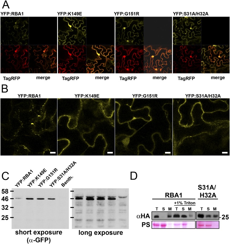
Fig. S5.
Localization of RBA1 (data related to Figs. 3 and 5). (A) Confocal imaging (magnification, 40×) of N. benthamiana-Agrobacteria transient expression of 35S YFP:RBA variants (yellow) and 35S TagRFP (red) in epidermal cells 24 h postinoculation (before visible symptoms). (B) Enlarged YFP images from the lower right quadrant of YFP images from A. (Scale bars, 40 uM.) (C) Western blot (anti-GFP) demonstrating that localization images reflect full-length fusion proteins. The short exposure demonstrates that mutants accumulate at higher levels. The long exposure shows no detectable free GFP, indicating that nuclear localization is reflective of full-length protein. Nonspecific plant cross-reacting bands indicate roughly equal loading among samples. (D) Microsomal fractionation of transiently overexpressed HA-RBA1 in N. benthamiana. M, microsomal pellet; S, soluble lysate; T, total lysate after centrifugation. Lanes are equally loaded with cell equivalents. PS, Ponceau stain to indicate protein loading of the membrane.

