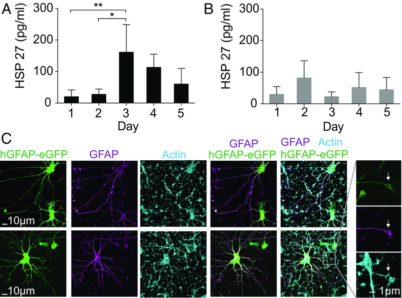
Fig. 5.
Longitudinal sampling from hiPSC-derived cardiomyocytes and astrocytes. (A and B) Longitudinal HSP27 extraction from the same hiPSC-CMs for 4 d. (A) The cardiomyocytes were stimulated by increasing the temperature to 44 °C for 30 min before sampling at day 2. An up-regulation of HSP27 was observed at day 3 (n = 4; *P < 0.05; Tukey’s post hoc test, one-way ANOVA). The HSP27 level started to drop at day 4. (B) Non–heat-shocked hiPSC-CMs were longitudinally sampled for 4 d (n = 4, P > 0.05; one-way ANOVA). (C) Representative images of astrocytes derived from hiPSCs in 3D cultures (hCS) and cultured in monolayer on NS. Astrocytes are labeled fluorescently with a lentiviral reporter (hGFAP::eGFP) and immunostained with an anti-GFAP antibody. The morphology of astrocytes was maintained even after 20 d of culture and repeated sampling on the NS platform. Arrowheads indicate structures that are approximately 1 µm in size, possibly NS that are surrounded by processes.

