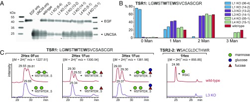
Fig. 2.
UNC5A secretion and C-mannosylation in single knockouts of DPY19 enzymes. (A) Secreted UNC5A in wild-type and single knockout cells. Purified UNC5A TSRs secreted from equal numbers of WT cells or two independent knockouts of DPY19L1 (L1 KO), DPY19L3 (L3 KO), and DPY19L4 (L4 KO) were analyzed using Western blotting with anti-myc antibody. A construct of myc-tagged EGF repeats 9–14 of mouse Notch1 was used as transfection and secretion control. (B) Quantification of extracted ion chromatograms of DPY19 knockouts. EIC profiles for the different glycoforms of TSR1 were quantified by integration and presented as percentage of the total TSR1 intensity. All peptides, independent of fucosylation (see Fig. 1B), were divided into species without or with one, two, or three mannoses. (C) Comparison of dimannosylated species of TSR1 and W3 mannosylation of TSR2 in wild-type and DPY19L3 (clone 14-1) knockout cells. MS/MS analysis of the dimannosylated TSR1 peptides with m/z = 1381.56 (SI Appendix, Fig. S4) confirmed that the DPY19L3 knockout completely lacks mannosylation of W3. All graphs are shown with equal intensities (13,000 counts per second).

