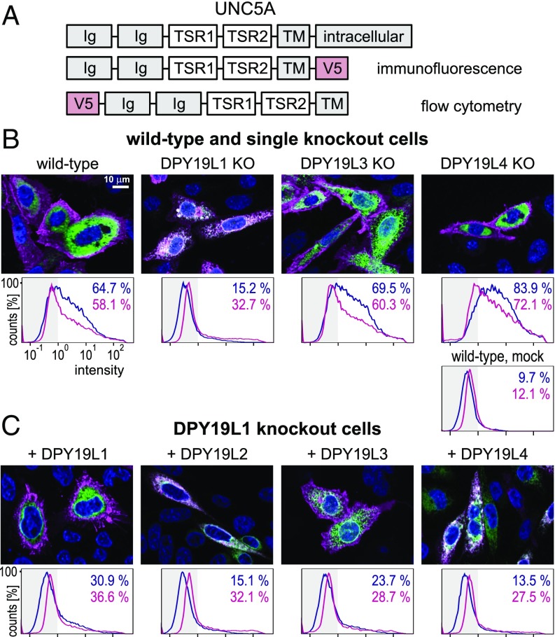
Fig. 4.
Subcellular localization of membrane-bound UNC5A. (A) UNC5A expression constructs. (B) Localization of UNC5A in knockout cells. (Upper) Immunofluorescence of transiently expressed C-terminal V5 tagged UNC5A construct (magenta) in wild-type CHO cells and in single knockouts of DPY19L1, -L3, and -L4. KDEL-tagged GFP (green) was used as an ER marker. (Lower) Flow cytometry of the same cells expressing N-terminally V5-tagged UNC5A. Cells were stained either alive on the cell surface by anti-V5 (blue line) or after fixation and permeabilization (magenta). Percentage of cells with a staining intensity above 100 [less than 1% in unstained cells and around 10% in mock transfected, but antibody-treated cells [mock WT)] are indicated in blue and magenta, respectively. (C) Effect of overexpression of DPY19 proteins in the DPY19L1 knockout. The four mouse DPY19 proteins were transiently expressed in the DPY19L1 knockout and immunostained as in B. (Scale bar, 10 μm.)

