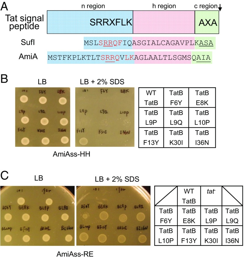
Fig. 1.
Isolation of signal sequence suppressors in tatB. (A) Schematic representation of a twin arginine signal peptide. The signal peptide sequences of E. coli Tat substrates SufI and AmiA are given underneath, with residues matching the Tat consensus motif in red, the consecutive arginines in red underline, and the signal peptidase cleavage site in black underline. (B and C) An example of screening results. Growth of MCDSSAC ΔtatABC coproducing the indicated TatB variants (with wild-type tatA and tatC) from pTAT101, alongside (B) the HH or (C) RE-substituted signal peptide variants of AmiA, on LB agar supplemented with chloramphenicol and kanamycin, with or without the addition of 2% (wt/vol) SDS as indicated. An 8-μL aliquot of each strain/plasmid combination following aerobic growth to an OD600 of 1.0 was spotted and incubated for 16 h at 37 °C.

