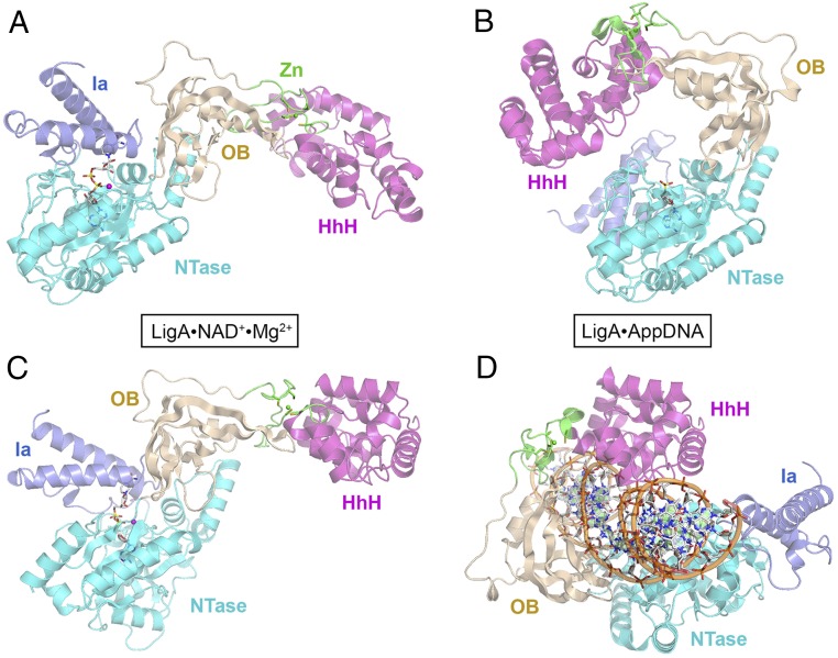
Fig. 2.
Structure of a LigA•NAD+ Michaelis complex and comparison with DNA-bound LigA. (A and B) The tertiary structures of EcoLigA-(K115M)•NAD+•Mg2+ (A, with ATP shown as a stick model) and EcoLigA•AppDNA (B, with AMP shown as a stick model, absent the nicked DNA) were aligned with respect to their NTase domains and then offset horizontally. (C and D) The structures of EcoLigA-(K115M)•NAD+•Mg2+ (C) and EcoLigA•AppDNA (D, with AppDNA) were aligned with respect to their HhH domains and then offset horizontally. The Ia (blue), NTase (cyan), OB (beige), Zn (green, with the Zn atom shown as a green sphere), and HhH (magenta) domains are color-coded.

