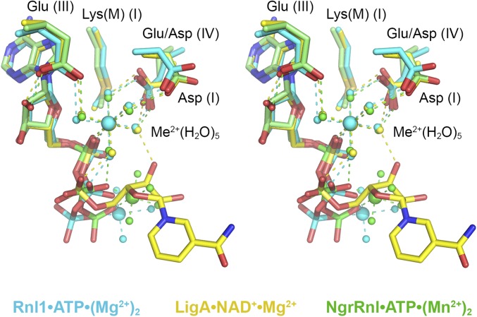
Fig. 4.
Comparison of the Michaelis complexes of ATP-dependent and NAD+-dependent polynucleotide ligases. Stereoview of the superimposed active sites of T4 Rnl1-(K99M)•ATP•(Mg2+)2 (with carbon atoms, waters, and Mg2+ colored cyan), EcoLigA-(K115M)•NAD+•Mg2+ (yellow), and the NgrRnl-(K170M)•ATP•(Mn2+)2 (green). Atomic contacts to the nucleotides and catalytic Me2+(H2O)5 complexes are shown as color-coded dashed lines.

