Fig. 2.
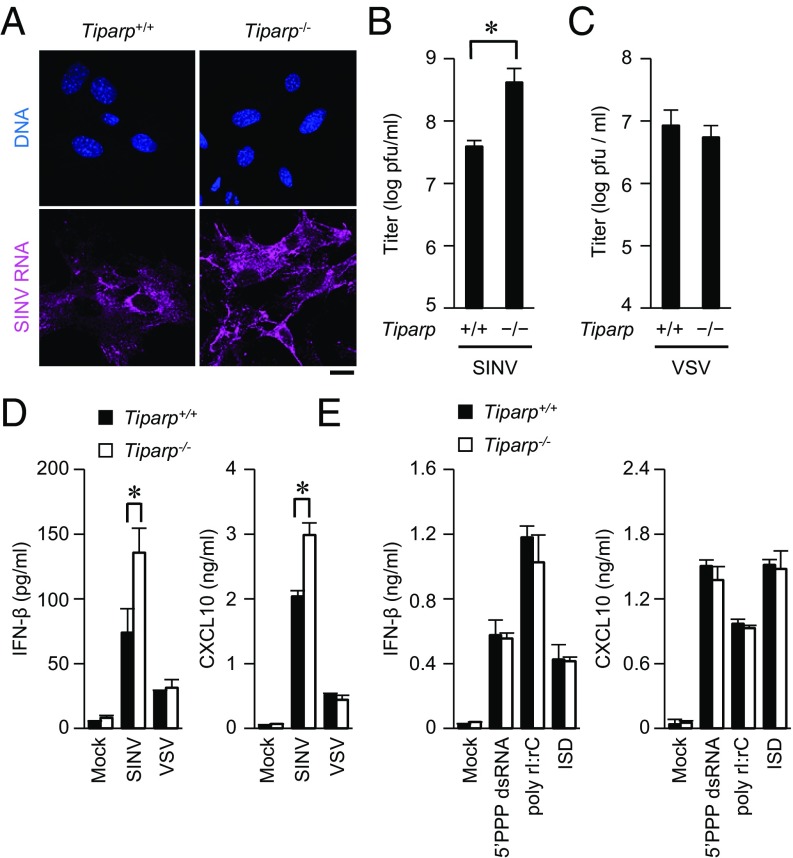
Loss of TIPARP enhances SINV replication. (A) Primary Tiparp+/+ and Tiparp−/− MEFs were infected with SINV (MOI = 1) for 12 h. Fixed samples were subjected to an RNA fluorescence in situ hybridization analysis of SINV RNA and to Hoechst 33342 staining of genomic DNA. (B and C) Primary Tiparp+/+ and Tiparp−/− MEFs were infected with SINV (MOI = 0.1) (B) or VSV (MOI = 0.1) (C) for 24 h. The viral titers in culture supernatants were determined by 50% tissue culture infectious dose assay. (D and E) ELISA of IFN-β and CXCL10 in culture supernatants of primary Tiparp+/+ and Tiparp−/− MEFs. (D) Cells were infected with SINV (MOI = 1) or VSV (MOI = 1) for 24 h. (E) Cells were stimulated with 5′PPP dsRNA (1 μg/mL), poly rI:rC (1 μg/mL), and IFN stimulatory DNA (ISD) (1 μg/mL), together with Lipofectamine 2000, for 24 h. (Scale bar, 20 μm.) Experiments were performed at least three times, and representative data are shown (means ± SD of three independent samples). *P < 0.05.

