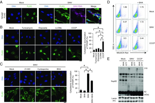
Fig. 5.
Cytoplasmic accumulation of TIPARP promotes elimination of SINV. (A) Wild-type MEFs stably expressing TIPARP-FLAG were infected with SINV (MOI = 5) for 12 h. Fixed samples were subjected to RNA fluorescence in situ hybridization analysis of SINV RNA, immunocytochemistry analysis of TIPARP-FLAG, and Hoechst 33342 staining of genomic DNA. (B and C) Wild-type MEFs stably expressing TIPARP-FLAG were treated with tunicamycin (5 μg/mL), etoposide (10 μM), Leu-Leu methyl ester hydrobromide (LLOMe) (100 μM), or CCCP (10 μM) for 6 h (B). Wild-type MEFs stably expressing TIPARP-FLAG were infected with SINV (MOI = 5), together with Z-VAD (10 μM), cyclosporin A (5 μM), or BHA (5 μM) for 12 h (C). Fixed samples were subjected to immunocytochemistry analysis of FLAG-tagged protein and Hoechst 33342 staining of genomic DNA. Frequencies of MEFs with cytoplasmic accumulation of TIPARP were determined. (D) Wild-type MEFs were infected with SINV (MOI = 5) or stimulated with CCCP (10 μM), with or without BHA (5 μM), for 12 h, and then stained with MitoSOX Red. Samples were subjected to flow cytometric analysis to measure the level of mitochondrial reactive oxygen species. SSC-A, side scatter area. (E) U373-CD14 cells were infected with SINV (MOI = 5) or stimulated with CCCP (50 μM) in the presence or absence of BHA (5 μM) for 24 h. Cellular extracts were treated with or without 100 mM DTT. Samples were suspended in 2-mercaptoethanol-free loading buffer and were subjected to immunoblot analysis of Nup62 and lamin A/C. (Scale bars, 20 μm.) The experiments were performed three times, and representative data are shown (means ± SD of three independent samples). *P < 0.05.

