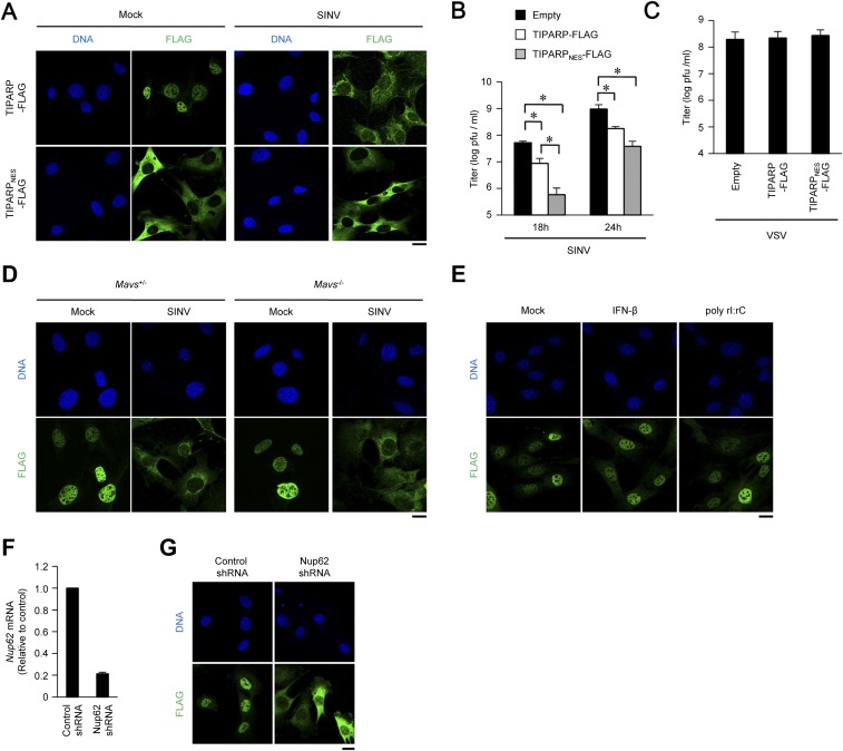
Fig. S5.
Cytoplasmic accumulation enhances anti-SINV activity of TIPARP. (A) Wild-type MEFs stably expressing TIPARP-FLAG and TIPARPNES-FLAG tagged protein were infected with SINV (MOI = 5) for 12 h. Fixed samples were subjected to immunocytochemistry analysis of FLAG-tagged protein and Hoechst 33342 staining of genomic DNA. (B and C) Wild-type MEFs stably expressing TIPARP-FLAG and TIPARPNES-FLAG-tagged protein were infected with SINV (MOI = 0.1) for the indicated durations (B) or VSV (MOI = 0.1) for 24 h (C). Viral titers in culture supernatants were determined by 50% tissue culture infectious dose assay. (D) Primary Mavs+/− or Mavs−/− MEFs stably expressing TIPARP-FLAG were infected with SINV (MOI = 5) for 12 h. Fixed samples were subjected to immunocytochemistry analysis of FLAG-tagged protein and Hoechst 33342 staining of genomic DNA. (E) Wild-type MEFs stably expressing TIPARP-FLAG were stimulated with IFN-β (10 ng/mL) or poly rI:rC (1 μg/mL) for 12 h. Fixed samples were subjected to immunocytochemistry analysis of FLAG-tagged protein and Hoechst 33342 staining of genomic DNA. (F) Wild-type MEFs were stably expressed with Nup62-specific and control shRNA. The levels of Nup62 mRNA were measured by quantitative reverse transcription-PCR. (G) Wild-type MEFs stably expressing TIPARP-FLAG with Nup62-specific shRNA or control shRNA. Fixed samples were subjected to immunocytochemistry analysis of TIPARP-FLAG and Hoechst 33342 staining of genomic DNA. (Scale bars, 20 μm.) Experiments were performed three times, and representative data are shown (means ± SD of three independent samples). *P < 0.05.

