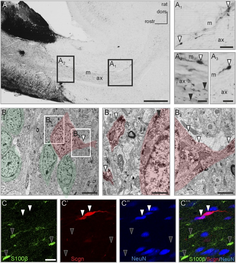
Fig. 1.
The presence and compartmentalization of secretagogin in rat RMS neurons. (A–A3) Secretagogin+ cells populate the rat RMS. In addition to somatic labeling (open arrowheads in A1–A3) a large number of individual processes were identified in the center of the middle domain of the stream (black arrowheads in A2; also Fig. S1E). Additionally, secretagogin+ cells often delineated the border of the RMS with their processes extended bipolarly (open arrowheads in A1 and A3). (B) Ultrastructural analysis identified secretagogin+ cells in close contact with chain-migrating neurons with secretagogin expression detectable both in the soma (black arrowhead in B1 and in processes arranged in parallel (open arrowheads in B1) or transversally (open arrowheads in B2). Chain-migrating neuronblasts and secretagogin+ cells are semitransparently color-coded in green and red, respectively. (C–C′′′) Secretagogin+ cells were invariably immunonegative for the gilal marker S100β (black arrowheads in C–C′′′) but were immunopositive for the neuron marker NeuN (white arrowheads in C′–C′′′). ax, axis of RMS; dors, dorsal; m, marginal zone of RMS; rostr, rostral; Scgn, secretagogin. (Scale bars: 500 µm in A, 30 µm in A1 and A2′; 20 µm in A3; 15 µm in C–C′′′; 10 µm in A2 and A4; 5 µm in B; 2 µm in B2; and 1 µm in B1.)

