Fig. 3.
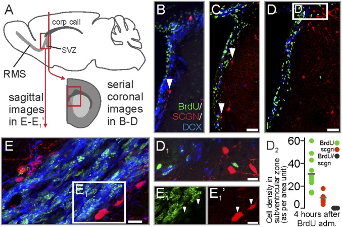
Secretagogin+ cells in the subventricular zone region and on entry into the RMS. (A) Schema of tissue sampling. (B–D2) Coronal sections. BrdU+ neuroblasts in the subventricular zone were demarcated toward the neighboring striatum by secretagogin+/BrdU− neurons. (E–E1′) Saggital sections. BrdU+ neuroblasts in the most proximal part of the RMS are demarcated by secretagogin+ neurons (white arrowheads in E1 and E1′). med, medial. (Scale bars: 80 µm in C and D; 10 µm in D1, E, and E1′.)

