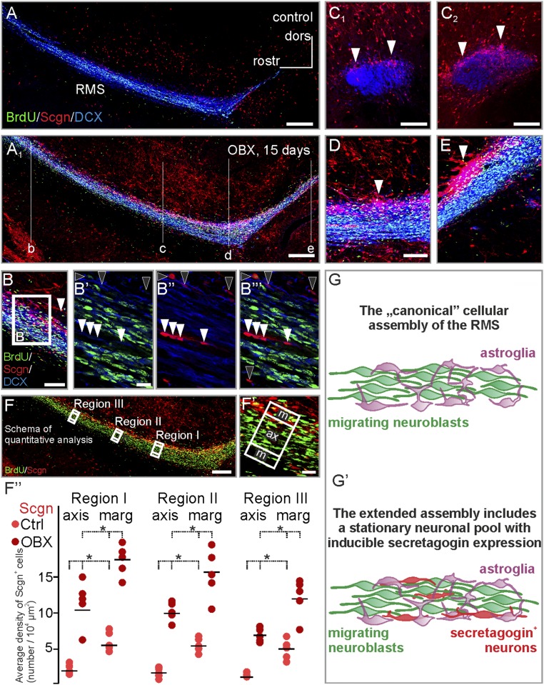
Fig. 4.
Unilateral bulbectomy increases the number of secretagogin-expressing cells in the rostral migratory stream. (A and A1) Secretagogin+ cells not only appeared in the otherwise secretagogin-immunonegative axis of the stream but also typically accumulated in its shell region 15 d after olfactory bulbectomy. Saggital sections are shown in B, D, and E. Coronal sections at the rostrocaudal level indicated in A1 are demonstrated in C1 and C2. (B′–B′′′) Olfactory bulbectomy increased the number of secretagogin+ neurons both in the marginal zone (black arrowheads) and in the axis (white arrowheads) of the RMS. (F–F′′) Fifteen days after bulbectomy, the number of secretagogin-expressing cells increased robustly in the axis and shell domains of the RMS compared with the number of cells detected in sham-operated controls. Secretagogin+ neurons and BrdU+ neuroblasts were counted manually, and their density was calculated in the marginal and axial domains (F′) of the distal RMS. To exclude region-specific differences, we carried out identical measurements in three regions of the distal RMS (F). The density secretagogin+ neurons increased significantly in both the marginal and axial domains of the RMS. (G and G′) In addition to astroglia and migrating neuroblasts, the rodent RMS contains secretagogin+ neurons (P < 0.05, Student’s t test). Images in A, A1, and F were acquired using the tile-and-stitch function. (Scale bars: 300 μm in A and A1; 200 μm in F; 100 μm in B, C1, C2, and D; and 10 μm in B′ and F′.)

