Fig. 7.
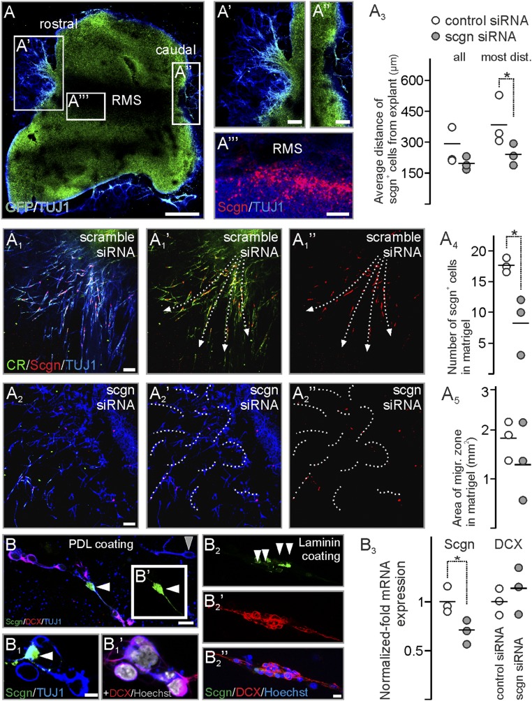
Secretagogin silencing disrupts neuroblast migration in vitro. (A–A′′′) RMS explants from P5 rat pups preserve polarity with migrating cells at the rostral (A′) but not caudal (A′′) pole with secretagogin+ neurons retaining their positions within explants (A′′′). (A1–A2′′) In controls neuroblasts migrate from the rostral pole of the RMS explants into Matrigel along corridors (arrows in A1′ and A1′′), which are shaped by secretagogin+ neurons. Secretagogin silencing disrupts cell migration (dashed curves in A2′ and A2′′). (A3–A5) Although the total area in the Matrigel occupied by migratory neurons decreased only nonsignificantly upon gene silencing, both the maximal and the average distance of secretagogin+ cells was significantly reduced from RMS exit. (B–B1′) In primary culture from P5 RMS plated on PDL, neuroblasts contacting secretagogin+ neurons (white arrowheads in B and B1) typically expressed DCX. (The gray arrowhead in B points to a DCX− neuron.) (B2–B2′′) On laminin-coated surfaces, secretagogin+ neurons (white arrowheads in B2) formed scaffolds for DCX+ neuroblasts. (B3) Secretagogin silencing decreased secretagogin mRNA levels but left DCX mRNA expression unaltered. P < 0.05, Student’s t test. (Scale bars: 500 µm in A; 70 µm in A′–A′′′; 25 µm in A1–A1′′; 10 µm in B; and 3 µm in B2′′.)

