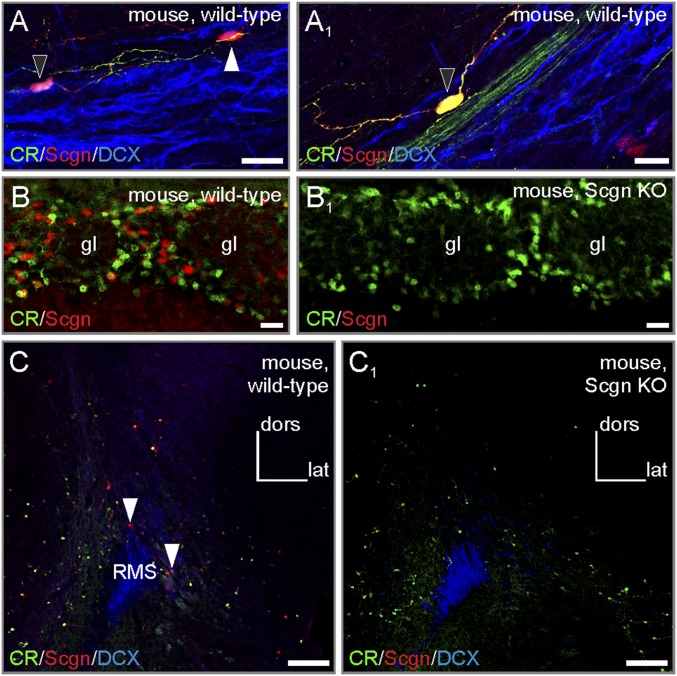
Fig. S3.
Secretagogin−/− mice demonstrate successful loss of function. (A and A1) Secretagogin+ neurons in wild-type RMSs typically expressed calretinin (black arrowheads). The white arrowhead points to a secretagogin+/calretinin− neuron). (B–C1) Secretagogin−/− mice lacked secretagogin expression in both the olfactory bulb (B and B1) and RMS (C and C1). (Scale bars: 100 µm in C and C1; 20 µm in B and B1; 10 µm in A and A1.)

