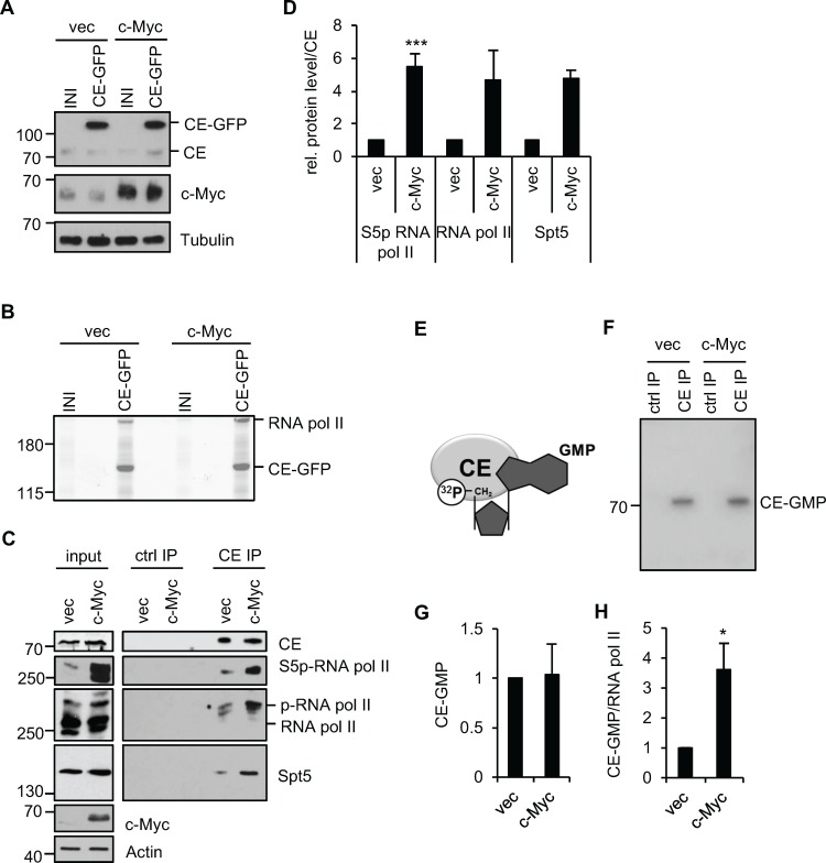
Figure 1. c-Myc increases the interaction of CE with RNA pol II.
(A) Protein extracts from IMEC/vec and IMEC/c-Myc, expressing CE-GFP or empty vector (INI) were analysed by Western blotting. (B) CE-GFP complexes were immunoprecipitated, resolved by SDS-PAGE and Coomassie-stained. Mass spectrometry was used to identify proteins. (C) CE complexes were immunoprecipitated from IMEC/vec and IMEC/c-Myc and analysed by Western blotting. FLAG antibody used as a control (ctrl IP). Input and immunoprecipitation (IP) panels are different exposures of the same Western blots. (D) ImageJ software was used to quantify Western blot signal for S5p-RNA pol II, RNA pol II pan and Spt5 in CE IP, normalised to CE. Error bars represent standard error of the mean, n=4 (S5p-RNA pol II) or n=2 (RNA Pol II and Spt5). (E) CE hydrolyses [α-32P] GTP, yielding a 32P-labelled CE-GMP intermediate, an approximation of CE activity. (F) CE IPs from nuclear extracts of IMEC/vec and IMEC/c-Myc were incubated with [α-32P]GTP, resolved by SDS-PAGE and 32P detected by phosphorimager. (G) Signal quantified by phosphoimager. Error bars represent standard error of the mean, n=5. (H) RNA pol II was immunoprecipitated from IMEC/vec and IMEC/c-Myc and 32P-GMP binding detected by phosphoimager. CE-32P-GMP signal normalised to RNA pol II (detected by Western blot). Error bars represent standard error of the mean, n=4. Significance relative to control calculated by Student's T-test; * = p≤0.05, *** = p≤0.001.

