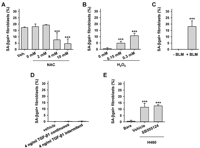
Figure 3. Effect of oxidative stress and exogenous TGF-β1 on fibroblast senescence induction by LCC cells.

A. Average percentage of SA-βgal+ CCD-19Lu fibroblasts co-cultured with H460 in the presence of increasing doses of the antioxidant NAC or vehicle. B, C. Average percentage of SA-βgal+ CCD-19Lu fibroblasts in response to direct or indirect oxidative stress elicited by (B) 2h treatment of H2O2 followed by 4 days of recovery or (C) 9 day treatment with bleomycin (BLM). D. Average percentage of SA-βgal+ CCD-19Lu fibroblasts daily treated with TGF-β1-continuously or intermitently for 4h/day as in [8]- for 2 weeks. E. Average percentage of SA-βgal+ CCD-19Lu fibroblasts co-cultured with H460 in the presence 5 μM of the TGF-β pathway inhibitor SB505124 for 9 days. All results correspond to two replicates from at least three independent experiments. All pair-wise comparisons were performed with respect to Bare or vehicle.
