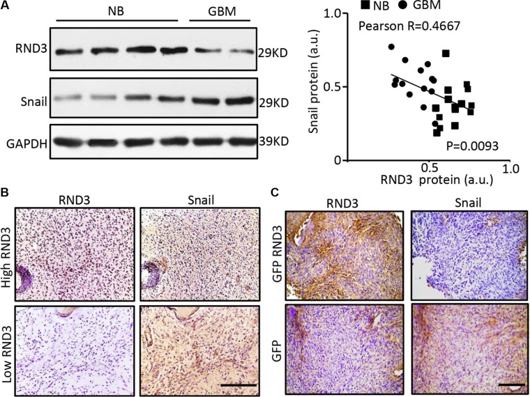
Figure 4. RND3 expression levels were inversely correlated with Snail1 expression levels in human and mouse GBM tissues.
(A–B) the protein levels of RND3 and Snail1 in human GBM tissues are detected by immunoblotting and immunohistochemical staining (brown). Quantification of the immunoblotting for RND3 and Snail showed an inverse correlation of the two protein expressions in human normal brain (NB) and glioblastoma (GBM) tissues (n = 30) (A, right panel). (C) Significant decrease in snail expression was shown in the GBM xenograft nude mouse brains with intracranial implantation of U251 cells overexpressing GFP-RND3 compared with the control mice with the implantation of U251 cells expressing GFP only. Pearson's test was performed for the correlation analysis. Scale bars represent 100 μm.

