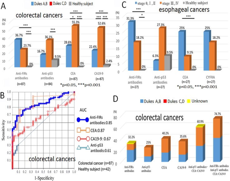
Figure 3. Diagnostic value of anti-FIRs antibodies in the sera of colorectal and esophageal cancer patients.
(A) The detection frequencies of anti-FIRs antibodies, His-tagged-FIRΔexon2 purified proteins as antigens, in the sera of early/advanced stages of colorectal cancer patients and healthy subjects were shown. (B) ROC (receiver operating characteristic) curve of colorectal cancer is indicated. The area under the curve (AUC) for anti-FIRs antibodies is 0.85, for anti-p53 antibody is 0.61, for CEA is 0.87 and for CA19-9 is 0.67. The best cutoff values for anti-FIRs antibodies, anti-p53 antibody, CEA and CA19-9 are 0.926%, 0.719 U/ml, 1.953 ng/ml and 12.8 U/ml, respectively. (C) The detection frequencies of anti-FIRs antibodies in the different stages of esophageal cancer patients and healthy subjects were also shown in graphical view. (D) The detection frequencies of anti-FIRs antibodies and three tumor markers in the different stages of colorectal cancer patients were shown in graphical view. The combination of three markers with anti-FIRs antibodies further increased the detection rate up to 74.7%.

