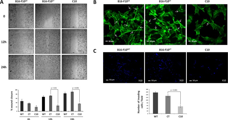
Figure 4. IGF-1 downregulation decreases cell invasion and migration.
(A) Scratchwound healing assay. Cells were grown to confluence and then used for wound healing migration assays. Artificial wounds were made in confluent monolayers of C10, B16-F10CT or B16-F10WT cells. Top, Migration of melanoma cells towards the wound, photographed after 6 h, 12 h and 24 h. The top panel shows one of three independent experiments. Bottom, Data are shown as means ± SEM. No significant differences in migratory activity were observed between B16-F10WT and B16-F10CT cells, with 67.7 ± 5.4% and 73.3 ± 2.4%, respectively, of the area displaying recovery at 12 h and 83.9 ± 6.3% and 91.6 ± 3.1%, respectively, of the area displaying recovery at 24 h. However, C10 clones displayed lower levels of migratory activity, with only 26.7 ± 5.5% (p < 0.01, n = 3) and 35.9 ± 1.4% (p < 0.001, n = 3) of recovery at 12 h and 24 h, respectively. (B) F-actin expression was analyzed by fluorescence microscopy with phalloidin (green) as a marker of membrane protrusions. The scale bar represents 10 μm. White arrowheads indicate the pseudopods. Each experiment was performed three times. (C) Invasion assay. C10, B16-F10CT and B16-F10WT cells were cultured in Matrigel Transwells. Their invasive potential was analyzed 96 h later, by fluorescence microscopy. The nuclei are stained blue with DAPI. Top, Representative images of three independent experiments are shown. The scale bar represents 50 μm. Bottom, Data are shown as means ± SEM. C10 clones have a lower invasion capacity (54.2 ± 9.2 cells per field) than parental (156.6 ± 8.3 cells per field) and control cells (131.6 ± 12.6 cells per field, p < 0.001, n = 3).

