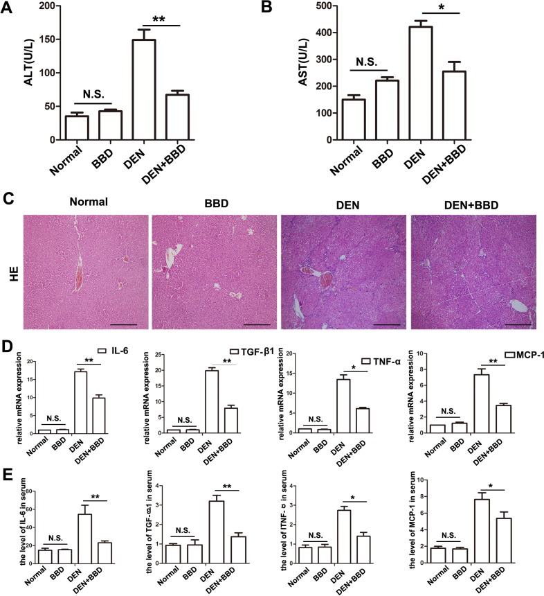
Figure 1. BBD ameliorated liver injury and inflammation in DEN-injured rats.
DEN was used to build a fibrotic model for studying the effect of BBD. Peripheral vein serum of each rat was collected at 8th week. Then, the levels of ALT (A) and AST (B) were measured to reflect liver injure. (C) HE staining (× 200) was performed to reflect hepatocyte injured and inflammatory cells infiltration. (D) The expression of inflammatory cytokines were abstracted from each fresh liver tissue and measured by RT-PCR. (E) The level of inflammatory cytokines in peripheral vein serum was also analyzed by ELISA. The unit is ng/L. Compared to DEN group, *P < 0.05; **P < 0.01; compared to Normal group, NS: no significance, by two-tailed Student's t test, n = 8, bar = 1 mm.

