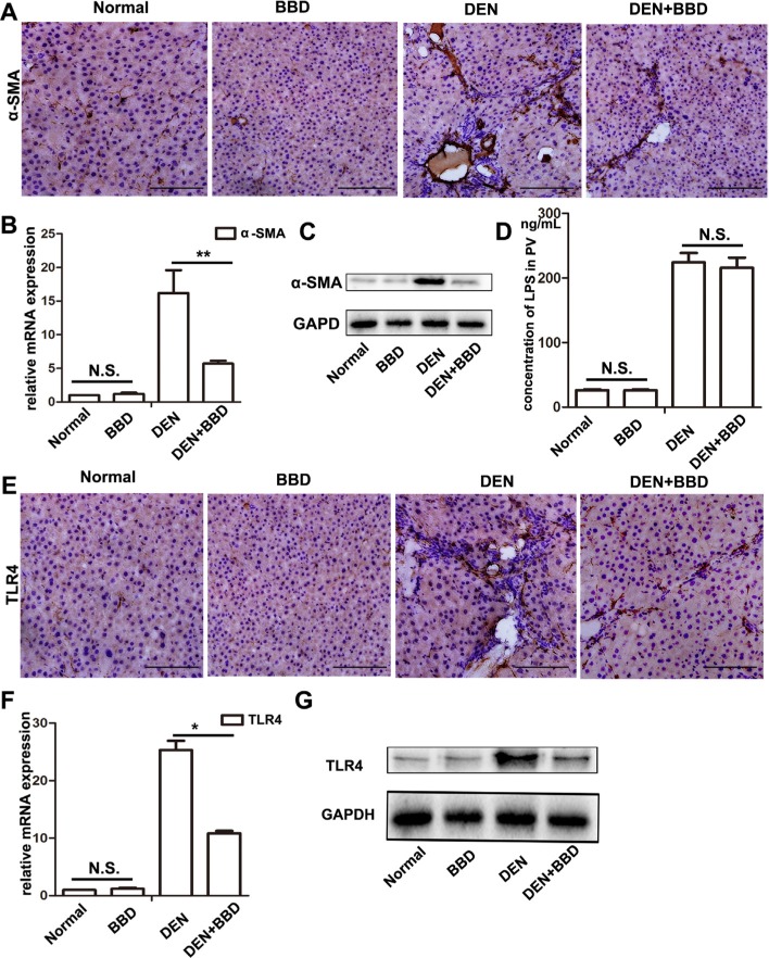
Figure 3. BBD inhibited hepatic stellate cells activation and the expression of TLR4 without attenuating the concentration of LPS.
4-μm-thick paraffin-embedded liver section was using for immunohistochemical staining (IHC). (A–C)The protein expression of α-SMA (× 200), a marker of activated HSCs, was analyzed by IHC, and quantitative measured by RT-PCR and Western blot. (D) Concentration of LPS in portal vein serum was detected by Rat endotoxin ElISA test kit. (E–G) The protein expression of TLR4 (× 200), a receptor specially bind to LPS, was determined by IHC, and also quantitative measured by RT-PCR and Western blot. Compared to DEN group, *P < 0.05; **P < 0.01, compared to Normal group, NS: no significance, by two-tailed Student's t test, n = 8, bar = 1 mm.

