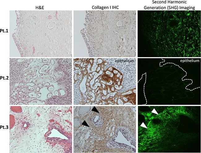
Figure 3. Representative images from individual NMIBC patients demonstrating Hematoxylin & Eosin (H&E) analysis, Collagen I protein expression by immunohistochemical analysis (IHC) and Second Harmonic Generation (SHG) imaging (20x serial sections from left to right).

Patient 1 (Pt.1) – Carcinoma-in-situ (CIS) with normal lamina propria (LP), collagen I staining by IHC and SHG imaging of curved collagen fibers, no progression. Patient 2 (Pt.2) – Papillary tumor with increased collagen I IHC staining within vascular stroma, also visualized on SHG, no progression. Patient 3 (Pt.3)– increased sub-epithelial LP staining and fibers with low curvature ratio by SHG imaging, experienced progression to MIBC. Corresponding areas of dense, straight-fibered collagen I deposition in the subepithelial stroma were marked with arrows.
