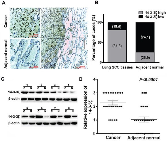
Figure 1. Expression of 14-3-3ζ is significantly up-regulated in lung SCC tissues compared with adjacent normal tissues by IHC and western blot.

A. IHC staining for 14-3-3ζ in human adjacent normal and lung SCC samples. Original magnification: 20× or 40×. High or low 14-3-3ζ expression was detected in cancerous or adjacent normal tissues, respectively. B. 14-3-3ζ expression patterns (high or low expression) were analyzed in human adjacent normal and lung SCC samples. C. Protein was extracted from matched lung SCC tissues and adjacent normal tissues and subjected to western blot analysis to examine 14-3-3ζ expression levels. β-Actin served as a loading control. D. The relative levels of 14-3-3ζ expression in lung SCC or adjacent normal tissue samples were quantified by western blot (1C) and normalized to β-actin (the internal control).
