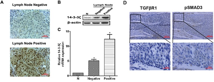
Figure 2. The expression of 14-3-3ζ, TGFβR1 and pSMAD3 proteins in lung SCC tissues and the relationship with clinicopathological characteristics.

A. The expression of 14-3-3ζ was evaluated in positive and negative lymph node metastasis by IHC. Original magnification: 40×. Representative images are shown. B. and C. Protein or RNA was extracted from lymph node metastasis of lung SCC patients or adjacent normal tissues and subjected to western blot or real-time PCR analysis to examine the level of 14-3-3ζ expression. β-Actin served as a loading control. All data are expressed as the means ± standard deviation (SD) for three independent experiments. N (adjacent normal tissue) served as a control. D. The expressions of TGFβR1 and pSMAD3 proteins were shown in the cytoplasm of cancer cells.
