
Figure 1. PRIMA-1Met inhibited the proliferation of CRC cells with different p53 status.
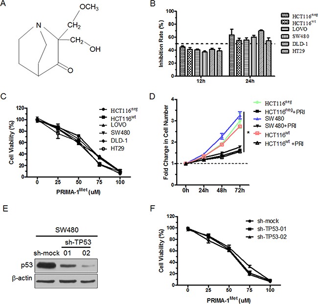
A. Chemical structure of PRIMA-1Met. B. CRC cells with different p53 status were treated with 25 uM PRIMA-1Met for 12 or 24 h. The cell viability was measured by MTS assay. C. Six colorectal cancer cells were exposed to PRIMA-1Met with various concentrations for 24 h and cell viability was measured by MTS assay. D. 1×10⁶ HCT116wt, HCT116neg or SW480 cells were treated with 25 uM PRIMA-1Met or control. Growth curve was measured by counting cell number at indicated time point. *P<0.05. PRI: PRIMA-1Met. E. SW480 cells were transiently transfected with sh-TP53 or sh-mock and p53 expression was analyzed by Western blot analysis. F. SW480 cells transfected with sh-mock or sh-TP53 were treated with various concentrations of PRIMA-1Met for 24 h and cell viability was measured by MTS assay. All data were shown as mean ± SEM from triplicate experiments.