
Figure 4. PRIMA-1Met specifically binds MEK in vitro and in vivo.
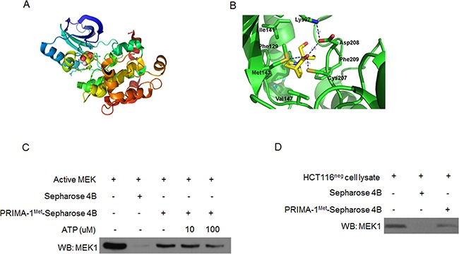
A. Computational binding model of PRIMA-1Met in proposed ATP-binding pocket of MEK1 monomer. PRIMA-1Met was shown as colored sphere and MEK1 monomer as cartoon. B. The proposed binding-position of hydrogen bonds between PRIMA-1Met and MEK1 was indicated. C. Pull-down assay was conducted in vitro by incubating 600 ug HCT116neg cell lysates with PRIMA-1Met-Sepharose 4B beads followed by Western blot analysis. D. In vivo ATP competitive binding assay was conducted by incubating 300 ng active MEK1 protein with 100 ul PRIMA-1Met-Sepharose 4B beads and various concentrations of ATP (0, 10, 100 uM) followed by Western blot analysis. Sepharose 4B only beads were used as negative control. Data shown were representative of three independent experiments.