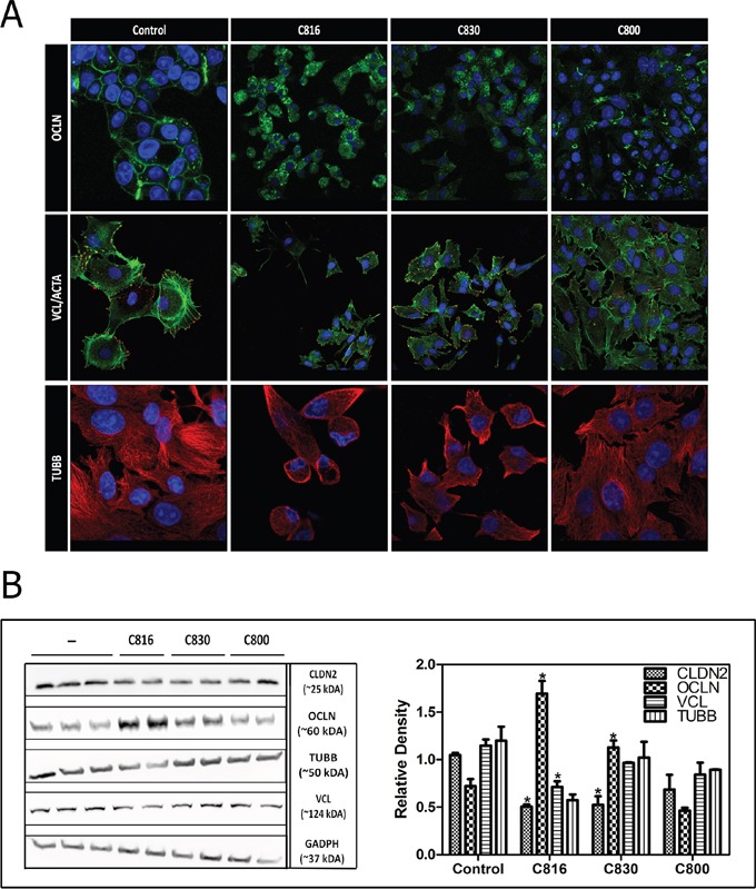
Figure 2. Crambescidins alter cell adherence and cytoskeletal integrity of tumor cells.

A. OCLN (green), VCL (red), F-ACTA (green) and β-TUBB (red) detection by confocal microscopy in control and 2.5 μM C816, C830 and C800-treated cells after 24 h. Colocalization of F-ACTA and VCL is shown in yellow and representative images of control and treated cells are shown. Hoechst 33258 was included for nuclei counterstaining (blue). B. Left: Determination of soluble CLDN2, OCLN, TUBB, VLC and GADPH levels. HepG2 cells were treated with 2.5 μM C816, C830 and C800 for 24 h and then soluble protein fractions obtained from cell lysates of treated and untreated cells were analyzed by western blot. Right: Quantification of the differences in protein levels among control and 2.5 μM C816, C830 and C800 treated cells.* Significant differences respect to controls (p <0.05).
