
Figure 1. Effect of ovarian cancer patient derived ascites on SKOV-3 cell migration and invasion.
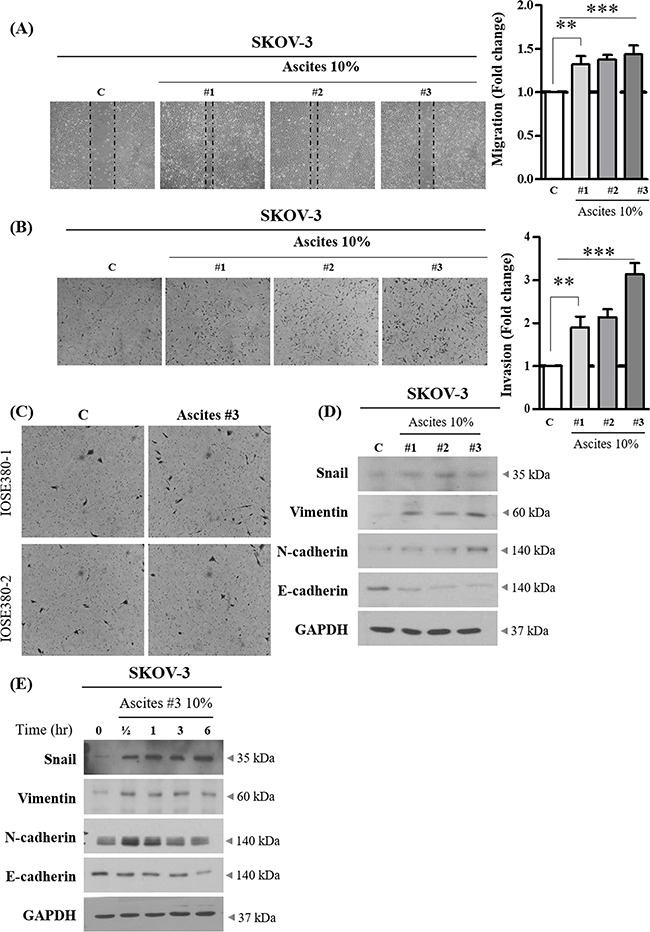
A. SKOV-3 cancer cells were treated with or without 10% ascites. After 24 hr, wound healing ability was verified by measuring wound closed area under a light microscope (magnification x 40). B. SKOV-3 cancer cells were seeded into the upper chamber of Matrigel-coated membrane in transwells. Cell invasion were induced with or without 10% ascites. After 24 hr, invaded cells at the bottom of the transwell were stained with 0.5% crystal violet and were counted under a light microscope (magnification x 200). C. IOSE380 cells were seeded into the upper chamber of Matrigel-coated membrane in transwells. Cell invasion were induced and counted as above. D. SKOV-3 cancer cells were treated with or without 10% ascites. After 24 hr, the expression levels of EMT molecular markers, Snail, Vimentin, N-cadherin and E-cadherin were examined by western blot. GAPDH was used as an internal control. E. SKOV-3 cancer cells were treated with or without 10% ascites for 0 – 6 hr. Total cell lysates were extracted and subjected to western blot as above. ** and *** represent P < 0.01 and P < 0.001, respectively.